Classification of bacteria according to their environment. Principles of classification of microorganisms
2.1. Systematics and nomenclature of microbes
The microbial world can be divided into cellular and non-cellular forms. Cellular forms of microbes are represented by bacteria, fungi and protozoa. They can be called microorganisms. Non-cellular forms are represented by viruses, viroids and prions.
The new classification of cellular microbes includes the following taxonomic units: domains, kingdoms, types, classes, orders, families, genera, species. The classification of microorganisms is based on their genetic relationship, as well as morphological, physiological, antigenic and molecular biological properties.
Viruses are often considered not as organisms, but as autonomous genetic structures, so they will be considered separately.
The cellular forms of microbes are divided into three domains. Domains Bacteria And Archaebacteria include microbes with a prokaryotic type of cell structure. Domain representatives Eukarya are eukaryotes. It consists of 4 kingdoms:
Mushroom kingdoms (Fungi, Eumycota);
kingdoms of protozoa (Protozoa);
kingdoms Chromista(chrome plates);
Microbes with unspecified taxonomic position (Microspora, microsporidia).
Differences in the organization of prokaryotic and eukaryotic cells are presented in table. 2.1.
Table 2.1. Signs of a prokaryotic and eukaryotic cell
2.2. Classification and morphology of bacteria
The term "bacteria" comes from the word bacteria, what does stick mean? Bacteria are prokaryotes. They are divided into two domains: Bacteria And Archaebacteria. Bacteria included in the domain Archaebacteria, represent one of the oldest forms of life. They have structural features of the cell wall (they lack peptidoglycan) and ribosomal RNA. There are no pathogens of infectious diseases among them.
Within a domain, bacteria are divided into the following taxonomic categories: class, phylum, order, family, genus, species. One of the main taxonomic categories is species. A species is a collection of individuals having the same origin and genotype, united by similar properties that distinguish them from other representatives of the genus. The species name corresponds to binary nomenclature, i.e. consists of two words. For example, the causative agent of diphtheria is written as Corynebacterium diphtheriae. The first word is the name of the genus and is written with a capital letter, the second word denotes the species and is written with a lowercase letter.
When a species is mentioned again, the generic name is abbreviated to its initial letter, e.g. C. diphtheriae.
A set of homogeneous microorganisms isolated on a nutrient medium, characterized by similar morphological, tinctorial (relation to dyes), cultural, biochemical and antigenic properties is called pure culture. A pure culture of microorganisms isolated from a specific source and different from other members of the species is called strain. Close to the concept of “strain” is the concept of “clone”. A clone is a collection of descendants grown from a single microbial cell.
To designate certain sets of microorganisms that differ in certain properties, the suffix “var” (variety) is used, therefore microorganisms, depending on the nature of the differences, are designated as morphovars (difference in morphology), resistant products (difference in resistance, for example, to antibiotics), serovars (difference in antigens), phagovars (difference in sensitivity to bacteriophages), biovars (difference in biological properties), chemovars (difference in biochemical properties), etc.
Previously, the basis for the classification of bacteria was the structural feature of the cell wall. The division of bacteria according to the structural features of the cell wall is associated with the possible variability of their coloring in one color or another using the Gram method. According to this method, proposed in 1884 by the Danish scientist H. Gram, depending on the staining results, bacteria are divided into gram-positive, stained blue-violet, and gram-negative, stained red.
Currently, the classification is based on the degree of genetic relatedness, based on studying the structure of the genome of ribosomal RNA (rRNA) (see Chapter 5), determining the percentage of guanine cytosine pairs (GC pairs) in the genome, constructing a restriction map of the genome, and studying the degree of hybridization. Phenotypic indicators are also taken into account: attitude to Gram staining, morphological, cultural and biochemical properties, antigenic structure.
Domain Bacteria includes 23 types, of which the following are of medical importance.
Most gram-negative bacteria are grouped into the phylum Proteobacteria(named after the Greek god Proteus, capable of taking on different forms). Type Proteobacteria divided into 5 classes:
Class Alphaproteobacteria(birth Rickettsia, Orientia, Erlichia, Bartonella, Brucella);
Class Betaproteobacteria(birth Bordetella, Burholderia, Neisseria, Spirillum);
Class Gammaproteobacteria(family representatives Enterobacteriaceae childbirth Francisella, Legionella, Coxiella, Pseudomonas, Vibrio);
Class Deltaproteobacteria(genus Bilophila);
Class Epsilonproteobacteria(birth Campilobacter, Helicobacter). Gram-negative bacteria also include the following types:
type Chlamydiae(birth Chlamydia, Chlamydophila), type Spirochaetes(birth Spirocheta, Borrelia, Treponema, Leptospira); type Bacteroides(birth Bacteroides, Prevotella, Porphyromonas).
Gram-positive bacteria come in the following types:
Type Firmicutes includes class Clostridium(birth Clostridium, Peptococcus), Class Bacilli (Listeria, Staphylococcus, Lactobacillus, Streptococcus) and class Mollicutes(birth Mycoplasma, Ureaplasma), which are bacteria that do not have a cell wall;
type Actinobacteria(birth Actinomyces, Micrococcus, Corynebacterium, Mycobacterium, Gardnerella, Bifidobacterium, Propionibacterium, Mobiluncus).
2.2.1. Morphological forms of bacteria
There are several main forms of bacteria: coccoid, rod-shaped, convoluted and branching (Fig. 2.1).
Spherical forms, or cocci- spherical bacteria 0.5-1 microns in size, which, according to their relative position, are divided into micrococci, diplococci, streptococci, tetracocci, sarcina and staphylococci.
Micrococci (from Greek. micros- small) - separately located cells.
Diplococci (from Greek. diploos- double), or paired cocci, are located in pairs (pneumococcus, gonococcus, meningococcus), since the cells do not separate after division. Pneumococcus (the causative agent of pneumonia) has a lanceolate shape on opposite sides, and gonococcus (the causative agent of gonorrhea) and meningococcus (the causative agent of
 Rice. 2.1. Shapes of bacteria
agent of epidemic meningitis) have the shape of coffee beans, with their concave surface facing each other.
Streptococci (from Greek. streptos- chain) - cells of a round or elongated shape, forming a chain due to cell division in the same plane and maintaining the connection between them at the site of division.
Sarcins (from lat. sarcina- bunch, bale) are arranged in the form of packets of 8 cocci or more, since they are formed during cell division in three mutually perpendicular planes.
Staphylococcus (from Greek. staphyle- grape bunch) - cocci located in the form of a bunch of grapes as a result of division in different planes.
Rod-shaped bacteria differ in size, shape of cell ends and relative position of cells. Cell length is 1-10 µm, thickness 0.5-2 µm. Sticks can be right
(Escherichia coli, etc.) and irregular club-shaped (Corynebacteria, etc.) shape. The smallest rod-shaped bacteria include rickettsia.
The ends of the rods can be cut off (anthrax bacillus), rounded (Escherichia coli), pointed (fusobacteria) or in the form of a thickening. In the latter case, the rod looks like a club (Corynebacterium diphtheria).
The slightly curved rods are called vibrios (Vibrio cholerae). Most rod-shaped bacteria are arranged randomly because the cells move apart after dividing. If, after division, the cells remain connected by common fragments of the cell wall and do not diverge, then they are located at an angle to each other (Corynebacterium diphtheria) or form a chain (anthrax bacillus).
Twisted Shapes- spiral-shaped bacteria, which come in two types: spirilla and spirochetes. Spirilla have the appearance of corkscrew-shaped convoluted cells with large curls. Pathogenic spirilla include the causative agent of sodoku (rat bite disease), as well as campylobacter and helicobacter, which have curves reminiscent of the wings of a flying seagull. Spirochetes are thin, long, convoluted bacteria that differ from spirilla in their smaller curls and pattern of movement. The peculiarities of their structure are described below.
Branching - rod-shaped bacteria, which can have Y-shaped branches found in bifidobacteria, can also be presented in the form of filamentous branched cells that can intertwine to form mycelium, as observed in actinomycetes.
2.2.2. Bacterial cell structure
The structure of bacteria has been well studied using electron microscopy of whole cells and their ultrathin sections, as well as other methods. The bacterial cell is surrounded by a membrane consisting of a cell wall and a cytoplasmic membrane. Under the shell there is protoplasm, consisting of cytoplasm with inclusions and a hereditary apparatus - an analogue of the nucleus, called the nucleoid (Fig. 2.2). There are additional structures: capsule, microcapsule, mucus, flagella, pili. Some bacteria are capable of forming spores under unfavorable conditions.
 Rice. 2.2. Structure of a bacterial cell: 1 - capsule; 2 - cell wall; 3 - cytoplasmic membrane; 4 - mesosomes; 5 - nucleoid; 6 - plasmid; 7 - ribosomes; 8 - inclusions; 9 - flagellum; 10 - pili (villi)
Cell wall- a strong, elastic structure that gives the bacterium a certain shape and, together with the underlying cytoplasmic membrane, restrains high osmotic pressure in the bacterial cell. It is involved in the process of cell division and transport of metabolites, has receptors for bacteriophages, bacteriocins and various substances. The thickest cell wall is found in gram-positive bacteria (Fig. 2.3). So, if the thickness of the cell wall of gram-negative bacteria is about 15-20 nm, then in gram-positive bacteria it can reach 50 nm or more.
The basis of the bacterial cell wall is peptidoglycan. Peptidoglycan is a polymer. It is represented by parallel polysaccharide glycan chains consisting of repeating N-acetylglucosamine and N-acetylmuramic acid residues connected by a glycosidic bond. This bond is broken by lysozyme, which is an acetylmuramidase.
A tetrapeptide is attached to N-acetylmuramic acid by covalent bonds. The tetrapeptide consists of L-alanine, which is linked to N-acetylmuramic acid; D-glutamine, which in gram-positive bacteria is combined with L-lysine, and in gram-tri-
 Rice. 2.3. Scheme of the architecture of the bacterial cell wall
beneficial bacteria - with diaminopimelic acid (DAP), which is a precursor of lysine in the process of bacterial biosynthesis of amino acids and is a unique compound present only in bacteria; The 4th amino acid is D-alanine (Fig. 2.4).
The cell wall of gram-positive bacteria contains small amounts of polysaccharides, lipids and proteins. The main component of the cell wall of these bacteria is multilayer peptidoglycan (murein, mucopeptide), accounting for 40-90% of the mass of the cell wall. Tetrapeptides of different layers of peptidoglycan in gram-positive bacteria are connected to each other by polypeptide chains of 5 glycine residues (pentaglycine), which gives the peptidoglycan a rigid geometric structure (Fig. 2.4, b). Covalently linked to the peptidoglycan of the cell wall of gram-positive bacteria teichoic acids(from Greek tekhos- wall), the molecules of which are chains of 8-50 glycerol and ribitol residues connected by phosphate bridges. The shape and strength of bacteria is given by the rigid fibrous structure of the multilayer peptidoglycan, with cross-links of peptides.
 Rice. 2.4. Structure of peptidoglycan: a - gram-negative bacteria; b - gram-positive bacteria
The ability of Gram-positive bacteria to retain gentian violet in combination with iodine when stained using Gram stain (blue-violet color of bacteria) is associated with the property of multilayer peptidoglycan to interact with the dye. In addition, subsequent treatment of a bacterial smear with alcohol causes a narrowing of the pores in the peptidoglycan and thereby retains the dye in the cell wall.
Gram-negative bacteria lose the dye after exposure to alcohol, which is due to a smaller amount of peptidoglycan (5-10% of the cell wall mass); they are discolored with alcohol, and when treated with fuchsin or safranin they become red. This is due to the structural features of the cell wall. Peptidoglycan in the cell wall of gram-negative bacteria is represented by 1-2 layers. The tetrapeptides of the layers are connected to each other by a direct peptide bond between the amino group of DAP of one tetrapeptide and the carboxyl group of D-alanine of the tetrapeptide of another layer (Fig. 2.4, a). Outside the peptidoglycan there is a layer lipoprotein, connected to peptidoglycan through DAP. Followed by outer membrane cell wall.
Outer membrane is a mosaic structure composed of lipopolysaccharides (LPS), phospholipids and proteins. Its inner layer is represented by phospholipids, and the outer layer contains LPS (Fig. 2.5). Thus, the outer mem-
 Rice. 2.5. Lipopolysaccharide structure
the brane is asymmetric. The outer membrane LPS consists of three fragments:
Lipid A has a conservative structure, almost the same in gram-negative bacteria. Lipid A consists of phosphorylated glucosamine disaccharide units to which long chains of fatty acids are attached (see Fig. 2.5);
Core, or core, crustal part (from lat. core- core), relatively conservative oligosaccharide structure;
A highly variable O-specific polysaccharide chain formed by repeating identical oligosaccharide sequences.
LPS is anchored in the outer membrane by lipid A, which causes LPS toxicity and is therefore identified with endotoxin. The destruction of bacteria by antibiotics leads to the release of large amounts of endotoxin, which can cause endotoxic shock in the patient. The core, or core part, of LPS extends from lipid A. The most constant part of the LPS core is ketodeoxyoctonic acid. O-specific polysaccharide chain extending from the core of the LPS molecule,
consisting of repeating oligosaccharide units, determines the serogroup, serovar (a type of bacteria detected using immune serum) of a particular strain of bacteria. Thus, the concept of LPS is associated with the concept of O-antigen, by which bacteria can be differentiated. Genetic changes can lead to defects, shortening of bacterial LPS and, as a result, the appearance of rough colonies of R-forms that lose O-antigen specificity.
Not all gram-negative bacteria have a complete O-specific polysaccharide chain, consisting of repeating oligosaccharide units. In particular, bacteria of the genus Neisseria have a short glycolipid called lipooligosaccharide (LOS). It is comparable to the R form, which has lost O-antigen specificity, observed in mutant rough strains E. coli. The structure of VOC resembles the structure of the glycosphingolipid of the human cytoplasmic membrane, so VOC mimics the microbe, allowing it to evade the host's immune response.
The matrix proteins of the outer membrane permeate it in such a way that protein molecules called porinami, border hydrophilic pores through which water and small hydrophilic molecules with a relative mass of up to 700 D pass.
Between the outer and cytoplasmic membrane is periplasmic space, or periplasm containing enzymes (proteases, lipases, phosphatases, nucleases, β-lactamases), as well as components of transport systems.
When the synthesis of the bacterial cell wall is disrupted under the influence of lysozyme, penicillin, protective factors of the body and other compounds, cells with an altered (often spherical) shape are formed: protoplasts- bacteria completely lacking a cell wall; spheroplasts- bacteria with a partially preserved cell wall. After removal of the cell wall inhibitor, such altered bacteria can reverse, i.e. acquire a full cell wall and restore its original shape.
Bacteria of the spheroid or protoplast type, which have lost the ability to synthesize peptidoglycan under the influence of antibiotics or other factors and are able to reproduce, are called L-shapes(from the name of the D. Lister Institute, where they first
have been studied). L-forms can also arise as a result of mutations. They are osmotically sensitive, spherical, flask-shaped cells of various sizes, including those passing through bacterial filters. Some L-forms (unstable), when the factor that led to changes in bacteria is removed, can reverse, returning to the original bacterial cell. L-forms can be produced by many pathogens of infectious diseases.
Cytoplasmic membrane in electron microscopy of ultrathin sections, it is a three-layer membrane (2 dark layers, each 2.5 nm thick, separated by a light intermediate one). In structure, it is similar to the plasmalemma of animal cells and consists of a double layer of lipids, mainly phospholipids, with embedded surface and integral proteins that seem to penetrate through the structure of the membrane. Some of them are permeases involved in the transport of substances. Unlike eukaryotic cells, the cytoplasmic membrane of a bacterial cell lacks sterols (with the exception of mycoplasmas).
The cytoplasmic membrane is a dynamic structure with mobile components, so it is thought of as a mobile fluid structure. It surrounds the outer part of the cytoplasm of bacteria and is involved in the regulation of osmotic pressure, transport of substances and energy metabolism of the cell (due to enzymes of the electron transport chain, adenosine triphosphatase - ATPase, etc.). With excessive growth (compared to the growth of the cell wall), the cytoplasmic membrane forms invaginates - invaginations in the form of complexly twisted membrane structures, called mesosomes. Less complexly twisted structures are called intracytoplasmic membranes. The role of mesosomes and intracytoplasmic membranes is not fully understood. It is even suggested that they are an artifact that occurs after preparing (fixing) a specimen for electron microscopy. Nevertheless, it is believed that derivatives of the cytoplasmic membrane participate in cell division, providing energy for the synthesis of the cell wall, and take part in the secretion of substances, sporulation, i.e. in processes with high energy consumption. Cytoplasm occupies the main volume of bacteria
cell and consists of soluble proteins, ribonucleic acids, inclusions and numerous small granules - ribosomes, responsible for the synthesis (translation) of proteins.
Ribosomes bacteria have a size of about 20 nm and a sedimentation coefficient of 70S, in contrast to the 80S ribosomes characteristic of eukaryotic cells. Therefore, some antibiotics, by binding to bacterial ribosomes, inhibit bacterial protein synthesis without affecting protein synthesis in eukaryotic cells. Bacterial ribosomes can dissociate into two subunits: 50S and 30S. rRNA is a conserved element of bacteria (“molecular clock” of evolution). 16S rRNA is part of the small ribosomal subunit, and 23S rRNA is part of the large ribosomal subunit. The study of 16S rRNA is the basis of gene systematics, allowing one to assess the degree of relatedness of organisms.
The cytoplasm contains various inclusions in the form of glycogen granules, polysaccharides, β-hydroxybutyric acid and polyphosphates (volutin). They accumulate when there is an excess of nutrients in the environment and act as reserve substances for nutrition and energy needs.
Volyutin has an affinity for basic dyes and is easily detected using special staining methods (for example, according to Neisser) in the form of metachromatic granules. With toluidine blue or methylene blue, volutin is stained red-violet, and the cytoplasm of the bacterium is stained blue. The characteristic arrangement of volutin granules is revealed in the diphtheria bacillus in the form of intensely stained cell poles. The metachromatic coloration of volutin is associated with a high content of polymerized inorganic polyphosphate. Under electron microscopy, they look like electron-dense granules 0.1-1 microns in size.
Nucleoid- equivalent to the nucleus in bacteria. It is located in the central zone of bacteria in the form of double-stranded DNA, tightly packed like a ball. The nucleoid of bacteria, unlike eukaryotes, does not have a nuclear envelope, nucleolus and basic proteins (histones). Most bacteria contain one chromosome, represented by a DNA molecule closed in a ring. But some bacteria have two ring-shaped chromosomes (V. cholerae) and linear chromosomes (see section 5.1.1). The nucleoid is revealed in a light microscope after staining with DNA-specific stains
methods: according to Feulgen or according to Romanovsky-Giemsa. In electron diffraction patterns of ultrathin sections of bacteria, the nucleoid appears as light zones with fibrillar, thread-like structures of DNA bound in certain areas to the cytoplasmic membrane or mesosome involved in chromosome replication.
In addition to the nucleoid, the bacterial cell contains extrachromosomal heredity factors - plasmids (see section 5.1.2), which are covalently closed rings of DNA.
Capsule, microcapsule, mucus.Capsule - a mucous structure more than 0.2 microns thick, firmly associated with the bacterial cell wall and having clearly defined external boundaries. The capsule is visible in imprint smears from pathological material. In pure bacterial cultures, the capsule is formed less frequently. It is detected using special methods of staining a smear according to Burri-Gins, which creates a negative contrast of the substances of the capsule: ink creates a dark background around the capsule. The capsule consists of polysaccharides (exopolysaccharides), sometimes of polypeptides, for example, in the anthrax bacillus it consists of polymers of D-glutamic acid. The capsule is hydrophilic and contains a large amount of water. It prevents the phagocytosis of bacteria. The capsule is antigenic: antibodies to the capsule cause its enlargement (capsule swelling reaction).
Many bacteria form microcapsule- mucous formation less than 0.2 microns thick, detectable only by electron microscopy.
It should be distinguished from a capsule slime - mucoid exopolysaccharides that do not have clear external boundaries. Mucus is soluble in water.
Mucoid exopolysaccharides are characteristic of mucoid strains of Pseudomonas aeruginosa, often found in the sputum of patients with cystic fibrosis. Bacterial exopolysaccharides are involved in adhesion (sticking to substrates); they are also called glycocalyx.
The capsule and mucus protect bacteria from damage and drying out, since, being hydrophilic, they bind water well and prevent the action of the protective factors of the macroorganism and bacteriophages.
Flagella bacteria determine the mobility of the bacterial cell. Flagella are thin filaments that take on
They originate from the cytoplasmic membrane and are longer than the cell itself. The thickness of the flagella is 12-20 nm, length 3-15 µm. They consist of three parts: a spiral filament, a hook and a basal body containing a rod with special discs (one pair of discs in gram-positive bacteria and two pairs in gram-negative bacteria). Flagella are attached to the cytoplasmic membrane and cell wall by discs. This creates the effect of an electric motor with a rod - a rotor - rotating the flagellum. The proton potential difference on the cytoplasmic membrane is used as an energy source. The rotation mechanism is provided by proton ATP synthetase. The rotation speed of the flagellum can reach 100 rps. If a bacterium has several flagella, they begin to rotate synchronously, intertwining into a single bundle, forming a kind of propeller.
Flagella are made of a protein called flagellin. (flagellum- flagellum), which is an antigen - the so-called H-antigen. Flagellin subunits are twisted in a spiral.
The number of flagella in different species of bacteria varies from one (monotrichus) in Vibrio cholerae to tens and hundreds extending along the perimeter of the bacterium (peritrichus), in Escherichia coli, Proteus, etc. Lophotrichs have a bundle of flagella at one end of the cell. Amphitrichy has one flagellum or a bundle of flagella at opposite ends of the cell.
Flagella are detected using electron microscopy of preparations coated with heavy metals, or in a light microscope after treatment with special methods based on etching and adsorption of various substances leading to an increase in the thickness of the flagella (for example, after silvering).
Villi, or pili (fimbriae)- thread-like formations, thinner and shorter (3-10 nm * 0.3-10 µm) than flagella. The pili extend from the cell surface and are composed of the protein pilin. Several types of pili are known. General type pili are responsible for attachment to the substrate, nutrition, and water-salt metabolism. They are numerous - several hundred per cell. Sex pili (1-3 per cell) create contact between cells, transferring genetic information between them by conjugation (see Chapter 5). Of particular interest are type IV pili, in which the ends are hydrophobic, as a result of which they curl; these pili are also called curls. Location
They are located at the poles of the cell. These pili are found in pathogenic bacteria. They have antigenic properties, bring bacteria into contact with the host cell, and participate in the formation of biofilm (see Chapter 3). Many pili are receptors for bacteriophages.
Disputes - a peculiar form of resting bacteria with a gram-positive type of cell wall structure. Spore-forming bacteria of the genus Bacillus, in which the size of the spore does not exceed the diameter of the cell are called bacilli. Spore-forming bacteria in which the size of the spore exceeds the diameter of the cell, which is why they take the shape of a spindle, are called clostridia, for example bacteria of the genus Clostridium(from lat. Clostridium- spindle). The spores are acid-resistant, therefore they are stained red using the Aujeszky method or the Ziehl-Neelsen method, and the vegetative cell is stained blue.
Sporulation, the shape and location of spores in a cell (vegetative) are a species property of bacteria, which allows them to be distinguished from each other. The shape of the spores can be oval or spherical, the location in the cell is terminal, i.e. at the end of the stick (in the causative agent of tetanus), subterminal - closer to the end of the stick (in the causative agents of botulism, gas gangrene) and central (in the anthrax bacillus).
The process of sporulation (sporulation) goes through a number of stages, during which part of the cytoplasm and chromosome of the bacterial vegetative cell are separated, surrounded by an ingrowing cytoplasmic membrane - a prospore is formed.
The prospore protoplast contains a nucleoid, a protein synthesizing system, and an energy production system based on glycolysis. Cytochromes are absent even in aerobes. Does not contain ATP, energy for germination is stored in the form of 3-glycerol phosphate.
The prospore is surrounded by two cytoplasmic membranes. The layer surrounding the inner membrane of the spore is called wall of spores, it consists of peptidoglycan and is the main source of cell wall during spore germination.
Between the outer membrane and the spore wall, a thick layer is formed consisting of peptidoglycan, which has many cross-links - cortex.
Located outside the outer cytoplasmic membrane spore shell, consisting of keratin-like proteins, co-
holding multiple intramolecular disulfide bonds. This shell provides resistance to chemical agents. The spores of some bacteria have an additional covering - exosporium lipoprotein nature. In this way, a multilayer, poorly permeable shell is formed.
Sporulation is accompanied by intensive consumption by the prospore and then by the developing spore shell of dipicolinic acid and calcium ions. The spore acquires heat resistance, which is associated with the presence of calcium dipicolinate in it.
The spore can persist for a long time due to the presence of a multilayer shell, calcium dipicolinate, low water content and sluggish metabolic processes. In soil, for example, the pathogens of anthrax and tetanus can persist for decades.
Under favorable conditions, spores germinate, going through three successive stages: activation, initiation, growth. In this case, one bacterium is formed from one spore. Activation is readiness for germination. At a temperature of 60-80 °C, the spore is activated for germination. Germination initiation lasts several minutes. The outgrowth stage is characterized by rapid growth, accompanied by destruction of the shell and emergence of the seedling.
2.2.3. Structural features of spirochetes, rickettsia, chlamydia, actinomycetes and mycoplasmas
Spirochetes- thin long convoluted bacteria. They consist of an outer membranous cell wall that surrounds a cytoplasmic cylinder. On top of the outer membrane there is a transparent cover of glycosaminoglycan nature. Under the outer membrane of the cell wall are fibrils that twist around the cytoplasmic cylinder, giving the bacteria a helical shape. The fibrils are attached to the ends of the cell and directed towards each other. The number and arrangement of fibrils varies among species. Fibrils are involved in the movement of spirochetes, giving the cells rotational, bending and translational motion. In this case, spirochetes form loops, curls, and bends, which are called secondary curls. Spirochetes do not perceive dyes well. They are usually painted according to Romanovsky-Giemsa or silver plated. Live
The form of a spirochete is examined using phase-contrast or dark-field microscopy.
Spirochetes are represented by three genera that are pathogenic for humans: Treponema, Borrelia, Leptospira.
Treponema(genus Treponema) have the appearance of thin, corkscrew-twisted threads with 8-12 uniform small curls. Around the protoplast of the treponema there are 3-4 fibrils (flagella). The cytoplasm contains cytoplasmic filaments. Pathogenic representatives are T. pallidum- the causative agent of syphilis, T. pertenue- causative agent of the tropical disease yaws. There are also saprophytes - inhabitants of the human oral cavity and the silt of reservoirs.
Borrelia(genus Borrelia), unlike treponemas, they are longer, have 3-8 large curls and 7-20 fibrils. These include the causative agent of relapsing fever (B. recurrentis) and pathogens of Lyme disease (V. burgdorferi) and other diseases.
Leptospira(genus Leptospira) They have shallow and frequent curls in the form of a twisted rope. The ends of these spirochetes are curved like hooks with thickenings at the ends. Forming secondary curls, they take on the shape of the letters S or C; have two axial fibrils. Pathogenic representative L. interrogans causes leptospirosis when ingested through water or food, leading to hemorrhage and jaundice.
Rickettsia have a metabolism independent of the host cell, however, it is possible that they receive high-energy compounds from the host cell for their reproduction. In smears and tissues they are stained according to Romanovsky-Giemsa, according to Macchiavello-Zdrodovsky (rickettsia are red, and infected cells are blue).
In humans, rickettsiae cause epidemic typhus. (R. prowazekii), tick-borne rickettsiosis (R. sibirica), Rocky Mountain spotted fever (R. rickettsii) and other rickettsioses.
The structure of their cell wall resembles that of gram-negative bacteria, although there are differences. It does not contain typical peptidoglycan: it completely lacks N-acetylmuramic acid. The cell wall consists of a double outer membrane, which includes lipopolysaccharide and proteins. Despite the absence of peptidoglycan, the cell wall of chlamydia is rigid. The cytoplasm of the cell is limited by the inner cytoplasmic membrane.
The main method for detecting chlamydia is Romanovsky-Giemsa staining. The color depends on the stage of the life cycle: elementary bodies appear purple against the background of the blue cytoplasm of the cell, reticular bodies appear blue.
In humans, chlamydia causes damage to the eyes (trachoma, conjunctivitis), urogenital tract, lungs, etc.
Actinomycetes- branching, filamentous or rod-shaped gram-positive bacteria. Its name (from Greek. actis- Ray, mykes- fungus) they received due to the formation in the affected tissues of drusen - granules of tightly intertwined threads in the form
rays extending from the center and ending in flask-shaped thickenings. Actinomycetes, like fungi, form mycelium - thread-like intertwining cells (hyphae). They form substrate mycelium, which is formed as a result of cell ingrowth into the nutrient medium, and aerial mycelium, which grows on the surface of the medium. Actinomycetes can divide by fragmentation of the mycelium into cells similar to rod-shaped and coccoid bacteria. On the aerial hyphae of actinomycetes, spores are formed that serve for reproduction. Actinomycete spores are usually not heat-resistant.
A common phylogenetic branch with actinomycetes is formed by the so-called nocardiform (nocardioform) actinomycetes - a collective group of rod-shaped bacteria of irregular shape. Their individual representatives form branching forms. These include bacteria of the genera Corynebacterium, Mycobacterium, Nocardia etc. Nocardi-like actinomycetes are distinguished by the presence in the cell wall of the sugars arabinose, galactose, as well as mycolic acids and large amounts of fatty acids. Mycolic acids and cell wall lipids determine the acid resistance of bacteria, in particular mycobacteria tuberculosis and leprosy (when stained according to Ziehl-Neelsen, they are red, and non-acid-resistant bacteria and tissue elements, sputum are blue).
Pathogenic actinomycetes cause actinomycosis, nocardia - nocardiosis, mycobacteria - tuberculosis and leprosy, corynebacteria - diphtheria. Saprophytic forms of actinomycetes and nocardiform actinomycetes are widespread in the soil, many of them are producers of antibiotics.
Mycoplasmas- small bacteria (0.15-1 µm), surrounded only by a cytoplasmic membrane containing sterols. They belong to the class Mollicutes. Due to the absence of a cell wall, mycoplasmas are osmotically sensitive. They have a variety of shapes: coccoid, filamentous, flask-shaped. These forms are visible under phase-contrast microscopy of pure cultures of mycoplasmas. On a dense nutrient medium, mycoplasmas form colonies that resemble fried eggs: a central opaque part immersed in the medium and a translucent periphery in the form of a circle.
Mycoplasmas cause atypical pneumonia in humans (Mycoplasma pneumoniae) and lesions of the genitourinary tract
(M. hominis and etc.). Mycoplasmas cause diseases not only in animals, but also in plants. Non-pathogenic representatives are also quite widespread.
2.3. Structure and classification of mushrooms
Mushrooms belong to the domain Eukarya, kingdom Fungi (Mycota, Mycetes). Recently, fungi and protozoa have been divided into separate kingdoms: kingdom Eumycota(real mushrooms), kingdom Chromista and kingdom Protozoa. Some microorganisms previously considered fungi or protozoa have been moved into a new kingdom Chromista(chrome plates). Fungi are multicellular or unicellular non-photosynthetic (chlorophyll-free) eukaryotic microorganisms with a thick cell wall. They have a nucleus with a nuclear envelope, cytoplasm with organelles, a cytoplasmic membrane and a multilayered rigid cell wall consisting of several types of polysaccharides (mannans, glucans, cellulose, chitin), as well as protein, lipids, etc. Some fungi form a capsule. The cytoplasmic membrane contains glycoproteins, phospholipids and ergosterols (as opposed to cholesterol, the main sterol of mammalian tissues). Most fungi are obligate or facultative aerobes.
Fungi are widespread in nature, especially in soil. Some mushrooms contribute to the production of bread, cheese, lactic acid products and alcohol. Other fungi produce antimicrobial antibiotics (eg, penicillin) and immunosuppressive drugs (eg, cyclosporine). Fungi are used by geneticists and molecular biologists to model various processes. Phytopathogenic fungi cause significant damage to agriculture, causing fungal diseases of cereal plants and grain. Infections caused by fungi are called mycoses. There are hyphal and yeast fungi.
Hyphal (mold) fungi, or hyphomycetes, consist of thin threads 2-50 microns thick, called hyphae, which are woven into a mycelium or mycelium (mold). The body of the mushroom is called the thallus. There are demacium (pigmented - brown or black) and hyaline (non-pigmented) hyphomycetes. Hyphae that grow into the nutrient substrate are responsible for feeding the fungus and are called vegetative hyphae. Hyphae, ra-
standing above the surface of the substrate are called aerial or reproductive hyphae (responsible for reproduction). Colonies have a fluffy appearance due to aerial mycelium.
There are lower and higher fungi: the hyphae of higher fungi are separated by partitions, or septa with holes. The hyphae of lower fungi do not have partitions, being multinucleated cells called coenocytic (from the Greek. koenos- single, common).
Yeast fungi (yeast) are mainly represented by individual oval cells with a diameter of 3-15 microns, and their colonies, unlike hyphal fungi, have a compact appearance. According to the type of sexual reproduction, they are distributed among higher fungi - ascomycetes and basidiomycetes. When reproducing asexually, yeast buds or divides. They can form pseudohyphae and false mycelium (pseudomycelium) in the form of chains of elongated cells - “sausages”. Fungi that are similar to yeast, but do not have a sexual method of reproduction, are called yeast-like. They reproduce only asexually - by budding or fission. The concepts of “yeast-like fungi” are often identified with the concept of “yeast”.
Many fungi have dimorphism - the ability to grow hyphal (mycelial) or yeast-like, depending on the cultivation conditions. In an infected organism, they grow in the form of yeast-like cells (yeast phase), and on nutrient media they form hyphae and mycelium. Dimorphism is associated with the temperature factor: at room temperature mycelium is formed, and at 37 ° C (at human body temperature) yeast-like cells are formed.
Fungi reproduce sexually or asexually. Sexual reproduction of fungi occurs with the formation of gametes, sexual spores and other sexual forms. Sexual forms are called teleomorphs.
Asexual reproduction of fungi occurs with the formation of corresponding forms called anamorphs. Such reproduction occurs by budding, fragmentation of hyphae and asexual spores. Endogenous spores (sporangiospores) mature inside a round structure - a sporangium. Exogenous spores (conidia) are formed at the tips of fruiting hyphae, the so-called conidiophores.
There are a variety of conidia. Arthroconidia (arthrospores), or talloconidia, are formed by uniform septation and dismemberment of hyphae, and blastoconidia are formed as a result of budding. Small unicellular conidia are called microconidia, large multicellular conidia are called macroconidia. Asexual forms of fungi also include chlamydoconidia, or chlamydospores (thick-walled large resting cells or a complex of small cells).
There are perfect and imperfect mushrooms. Perfect mushrooms have a sexual method of reproduction; these include zygomycetes (Zygomycota), ascomycetes (Ascomycota) and basidiomycetes (Basidiomycota). Imperfect fungi have only asexual reproduction; These include the formal conventional type/group of fungi - deuteromycetes (Deiteromycota).
Zygomycetes belong to the lower fungi (nonseptate mycelium). They include representatives of the genera Mucor, Rhizopus, Rhizomucor, Absidia, Basidiobolus, Conidiobolus. Distributed in soil and air. They can cause zygomycosis (mucoromycosis) of the lungs, brain and other human organs.
During asexual reproduction of zygomycetes, a sporangium is formed on the fruiting hypha (sporangiophores) - a spherical thickening with a shell containing numerous sporangiospores (Fig. 2.6, 2.7). Sexual reproduction in zygomycetes occurs with the help of zygospores.
Ascomycetes (marsupial fungi) have septate mycelium (except for unicellular yeasts). They got their name from the main fruiting organ - the bursa, or ascus, containing 4 or 8 haploid sexual spores (ascospores).
Ascomycetes include individual representatives (teleomorphs) of the genera Aspergillus And Penicillium. Most fungi genera Aspergillus, Penicillium are anamorphic, i.e. they reproduce only helplessly
 Rice. 2.6. Mushrooms of the genus Mucor(drawing by A.S. Bykov)
 Rice. 2.7. Mushrooms of the genus Rhizopus. Development of sporangium, sporangiospores and rhizoids
directly with the help of asexual spores - conidia (Fig. 2.8, 2.9) and should be classified according to this characteristic as imperfect fungi. In mushrooms of the genus Aspergillus at the ends of the fruiting hyphae, conidiophores, there are thickenings - sterigmata, phialides, on which chains of conidia are formed (“water mold”).
In mushrooms of the genus Penicillium(tassel) the fruiting hypha resembles a brush, since thickenings are formed from it (on the conidiophore), branching into smaller structures - sterigmata, phialides, on which there are chains of conidia. Some species of Aspergillus can cause aspergillosis and aflatoxicosis, and Penicillium can cause penicilliosis.
Representatives of ascomycetes are teleomorphs of the genera Trichophyton, Microsporum, Histoplasma, Blastomyces, as well as tremors
 Rice. 2.8. Mushrooms of the genus Penicillium. Chains of conidia extend from the phialids
 Rice. 2.9. Mushrooms of the genus Aspergillus fumigatus. Chains of conidia extend from the phialids
Basidiomycetes include cap mushrooms. They have septate mycelium and form sexual spores - basidiospores by detaching from the basidium - the terminal cell of the mycelium, homologous to the ascus. Basidiomycetes include some yeasts, such as teleomorphs Cryptococcus neoformans.
Deuteromycetes are imperfect fungi (Fungi imperfecti, anamorphic fungi, conidial fungi). This is a conditional, formal taxon of fungi, uniting fungi that do not have sexual reproduction. Recently, instead of the term “deuteromycetes”, the term “mitosporous fungi” was proposed - fungi that reproduce by non-sexual spores, i.e. by mitosis. When the fact of sexual reproduction of imperfect fungi is established, they are transferred to one of the known types - Ascomycota or Basidiomycota assigning the name to a teleomorphic form. Deuteromycetes have septate mycelium and reproduce only through the asexual formation of conidia. Deuteromycetes include imperfect yeasts (yeast-like fungi), for example, some fungi of the genus Candida, affecting the skin, mucous membranes and internal organs (candidiasis). They are oval in shape, 2-5 microns in diameter, divide by budding, form pseudohyphae (pseudomycelium) in the form of chains of elongated cells, and sometimes form hyphae. For Candida albicans the formation of chlamydospores is characteristic (Fig. 2.10). Deuteromycetes also include other fungi that do not have a sexual method of reproduction, belonging to the genera Epidermophyton, Coccidioides, Paracoccidioides, Sporothrix, Aspergillus, Phialophora, Fonsecaea, Exophiala, Cladophialophora, Bipolaris, Exerohilum, Wangiella, Alrernaria and etc.
 Rice. 2.10. Mushrooms of the genus Candida albicans(drawing by A.S. Bykov)
2.4. Structure and classification of protozoa
The simplest belong to the domain Eukarya, animal kingdom (Animalia), sub-kingdom Protozoa. It has recently been proposed to allocate protozoa to the rank of kingdom Protozoa.
The protozoan cell is surrounded by a membrane (pellicle) - an analogue of the cytoplasmic membrane of animal cells. It has a nucleus with a nuclear envelope and nucleolus, a cytoplasm containing the endoplasmic reticulum, mitochondria, lysosomes and ribosomes. The sizes of protozoa range from 2 to 100 microns. When stained according to Romanovsky-Giemsa, the nucleus of the protozoa is red, and the cytoplasm is blue. Protozoa move with the help of flagella, cilia or pseudopodia, some of them have digestive and contractile (excretory) vacuoles. They can feed as a result of phagocytosis or the formation of special structures. Based on the type of nutrition, they are divided into heterotrophs and autotrophs. Many protozoa (dysenteric amoeba, Giardia, Trichomonas, Leishmania, Balantidia) can grow on nutrient media containing native proteins and amino acids. Cell cultures, chicken embryos and laboratory animals are also used for their cultivation.
Protozoa reproduce asexually - by double or multiple (schizogony) division, and some also sexually (sporogony). Some protozoa reproduce extracellularly (Giardia), while others reproduce intracellularly (Plasmodium, Toxoplasma, Leishmania). The life cycle of protozoa is characterized by stages - the formation of the trophozoite stage and the cyst stage. Cysts are dormant stages, resistant to changes in temperature and humidity. Cysts are acid resistant Sarcocystis, Cryptosporidium And Isospora.
Previously, protozoa that cause diseases in humans were represented by 4 types 1 ( Sarcomastigophora, Apicomplexa, Ciliophora, Microspora). These types have recently been reclassified into larger numbers, and new kingdoms have emerged - Protozoa And Chromista(Table 2.2). To a new kingdom Chromista(chromovics) included some protozoa and fungi (blastocysts, oomycetes and Rhinosporidium seeberi). Kingdom Protozoa includes amoebas, flagellates, sporozoans and ciliates. They are divided into different types, among which are amoebas, flagellates, sporozoans and ciliates.
Table 2.2. Representatives of the kingdoms Protozoa And Chromista, of medical importance
 1 Type Sarcomastigophora consisted of subtypes Sarcodina And Mastigophora. Subtype Sarcodina(sarcodaceae) included the dysenteric amoeba, and the subtype Mastigophora(flagellates) - trypanosomes, leishmania, lamblia and trichomonas. Type Apicomplexa included class Sporozoa(spores), which included plasmodia of malaria, toxoplasma, cryptosporidium, etc. Type Ciliophora includes balantidia, and type Microspora- microsporidia.
End of table. 2.2
 Amoebas include the causative agent of human amoebiasis - amoebic dysentery (Entamoeba histolytica), free-living and non-pathogenic amoebas (intestinal amoeba, etc.). Amoebas reproduce binary asexually. Their life cycle consists of a trophozoite stage (a growing, motile cell, weakly stable) and a cyst stage. Trophozoites move with the help of pseudopodia, which capture and immerse nutrients into the cytoplasm. From
The trophozoite forms a cyst that is resistant to external factors. Once in the intestine, it turns into a trophozoite.
Flagellates are characterized by the presence of flagella: Leishmania has one flagellum, Trichomonas has 4 free flagella and one flagellum connected to a short undulating membrane. They are:
Flagellates of blood and tissues (Leishmania - the causative agents of leishmaniasis; Trypanosomes - the causative agents of sleeping sickness and Chagas disease);
Intestinal flagellates (giardia - the causative agent of giardiasis);
Flagellates of the genitourinary tract (Trichomonas vaginalis - the causative agent of trichomoniasis).
The ciliated ones are represented by balantidia, which affect the human colon (balantidiasis dysentery). Balantidia have a trophozoite and cyst stage. The trophozoite is mobile, has numerous cilia, thinner and shorter than flagella.
2.5. Structure and classification of viruses
Viruses are the smallest microbes belonging to the kingdom Virae(from lat. virus- I). They do not have a cellular structure and consist
The structure of viruses, due to their small size, is studied using electron microscopy of both virions and their ultrathin sections. The sizes of viruses (virions) are determined directly using electron microscopy or indirectly by ultrafiltration through filters with a known pore diameter, or by ultracentrifugation. The size of viruses ranges from 15 to 400 nm (1 nm is equal to 1/1000 μm): small viruses, the size of which is similar to the size of ribosomes, include parvoviruses and poliovirus, and the largest are variola virus (350 nm). Viruses differ in the shape of their virions, which have the form of rods (tobacco mosaic virus), bullets (rabies virus), spheres (poliomyelitis viruses, HIV), threads (filoviruses), sperm (many bacteriophages).
Viruses amaze the imagination with their diversity of structure and properties. Unlike cellular genomes, which contain uniform double-stranded DNA, viral genomes are extremely diverse. There are DNA and RNA viruses that are haploid, i.e. have one set of genes. Only retroviruses have a diploid genome. The genome of viruses contains from 6 to 200 genes and is represented by various types of nucleic acids: double-stranded, single-stranded, linear, circular, fragmented.
Among single-stranded RNA viruses, a distinction is made between genomic plus-strand RNA and minus-strand RNA (RNA polarity). The plus strand (positive strand) of the RNA of these viruses, in addition to the genomic (hereditary) function, performs the function of informational or messenger RNA (mRNA, or mRNA); it is a matrix for protein synthesis on the ribosomes of the infected cell. Plus-strand RNA is infectious: when introduced into sensitive cells, it can cause an infectious process.
cess. The minus strand (negative strand) of RNA viruses performs only a hereditary function; For protein synthesis, a complementary strand is synthesized on the minus strand of RNA. Some viruses have an ambipolar RNA genome (ambisense from Greek ambi- on both sides, double complementarity), i.e. contains plus and minus RNA segments.
There are simple viruses (for example, hepatitis A virus) and complex viruses (for example, influenza viruses, herpes, coronaviruses).
Simple, or non-enveloped, viruses have only nucleic acid associated with a protein structure called a capsid (from the Latin. capsa- case). Proteins associated with nucleic acid are known as nucleoproteins, and the association of viral capsid proteins with viral nucleic acid is called nucleocapsid. Some simple viruses can form crystals (eg foot-and-mouth disease virus).
The capsid includes repeating morphological subunits - capsomeres, composed of several polypeptides. The nucleic acid of the virion binds to the capsid to form a nucleocapsid. The capsid protects the nucleic acid from degradation. In simple viruses, the capsid is involved in attachment (adsorption) to the host cell. Simple viruses leave the cell as a result of its destruction (lysis).
Complex, or enveloped, viruses (Fig. 2.11), in addition to the capsid, have a membrane double lipoprotein envelope (synonym: supercapsid, or peplos), which is acquired by budding of the virion through the cell membrane, for example through the plasma membrane, nuclear membrane or endoplasmic reticulum membrane. The virus envelope contains glycoprotein spikes,
or spines, peplomers. Destruction of the shell with ether and other solvents inactivates complex viruses. Under the shell of some viruses is a matrix protein (M protein).
Virions have a helical, icosahedral (cubic) or complex type of capsid (nucleocapsid) symmetry. The helical type of symmetry is due to the helical structure of the nucleocapsid (for example, in influenza viruses, coronaviruses): capsomeres are arranged in a spiral along with the nucleic acid. The icosahedral type of symmetry is due to the formation of an isometrically hollow body from the capsid containing the viral nucleic acid (for example, in the herpes virus).
The capsid and shell (supercapsid) protect virions from environmental influences and determine selective interaction (adsorption) of their receptor proteins with certain
 Rice. 2.11. The structure of enveloped viruses with an icosahedral (a) and helical (b) capsid
cells, as well as antigenic and immunogenic properties of virions.
The internal structures of viruses are called the core. In adenoviruses, the core consists of histone-like proteins associated with DNA, in reoviruses - from proteins of the internal capsid.
Nobel Prize winner D. Baltimore proposed the Baltimore classification system based on the mechanism of mRNA synthesis. This classification places viruses in 7 groups (Table 2.3). International Committee on Taxonomy of Viruses (ICTV) adopted a universal classification system that uses taxonomic categories such as family (name ending in viridae), subfamily (name ends in virinae), genus (name ends in virus). The virus species did not receive a binomial name, like bacteria. Viruses are classified by the type of nucleic acid (DNA or RNA), its structure and the number of strands. They have double-stranded or single-stranded nucleic acids; positive (+), negative (-) nucleic acid polarity or mixed nucleic acid polarity, ambipolar (+, -); linear or circular nucleic acid; fragmented or non-fragmented nucleic acid. The size and morphology of virions, the number of capsomeres and the type of symmetry of the nucleocapsid, the presence of an envelope (supercapsid), sensitivity to ether and deoxycholate, place of reproduction in the cell, antigenic properties, etc. are also taken into account.
Table 2.3. Major viruses of medical importance
 Continuation of the table. 2.3
 End of table. 2.3
 Viruses infect animals, bacteria, fungi and plants. Being the main causative agents of human infectious diseases, viruses are also involved in the processes of carcinogenesis and can be transmitted in various ways, including through the placenta (rubella virus, cytomegalovirus, etc.), affecting the human fetus. They can also lead to post-infectious complications - the development of myocarditis, pancreatitis, immunodeficiency, etc.
In addition to viruses, non-cellular life forms include prions and viroids. Viroids are small molecules of circular, supercoiled RNA that do not contain protein and cause plant diseases. Pathological prions are infectious protein particles that cause special conformational diseases as a result of changes in the structure of normal cellular prion protein ( PrP c), which is present in the body of animals and humans. PrP with performs regulatory functions. It is encoded by the normal prion gene (PrP gene), located on the short arm of human chromosome 20. Prion diseases occur as transmissible spongiform encephalopathies (Creutzfeldt-Jakob disease, kuru, etc.). In this case, the prion protein acquires a different, infectious form, designated as PrP sc(sc from scrapie- scrapie is a prion infection of sheep and goats). This infectious prion protein has the appearance of fibrils and differs from normal prion protein in its tertiary or quaternary structure.
Tasks for self-preparation (self-control)
A. Label the microbes that are prokaryotes:
2. Viruses.
3. Bacteria.
4. Prions.
B. Note the distinctive features of a prokaryotic cell:
1. 70S ribosomes.
2. The presence of peptidoglycan in the cell wall.
3. Presence of mitochondria.
4. Diploid set of genes.
IN. Label the components of peptidoglycan:
1. Teichoic acids.
2. N-acetylglucosamine.
3. Lipopolysaride.
4. Tetrapeptide.
G. Note the structural features of the cell wall of gram-negative bacteria:
1. Mesodiaminopimelic acid.
2. Teichoic acids.
4. Porin proteins.
D. Name the functions of spores in bacteria:
1. Preservation of the species.
2. Heat resistance.
3. Dispersal of the substrate.
4. Reproduction.
1. Rickettsia.
2. Actinomycetes.
3. Spirochetes.
4. Chlamydia.
AND. Name the features of actinomycetes:
1. They have thermolabile spores.
2. Gram-positive bacteria.
3. There is no cell wall.
4. They have a convoluted shape.
Z. Name the features of spirochetes:
1. Gram-negative bacteria.
2. They have a motor fibrillary apparatus.
3. They have a convoluted shape.
AND. Name the protozoa that have an apical complex that allows them to penetrate into the cell:
1. Malarial plasmodium.
3. Toxoplasma.
4. Cryptosporidium.
TO. Name the distinctive feature of complex viruses:
1. Two types of nucleic acid.
2. The presence of a lipid membrane.
3. Double capsid.
4. Presence of non-structural proteins. L. Mark the higher mushrooms:
1. Mucor.
2. Candida.
3. Penicillium.
4. Aspergillus.
Microorganisms (microbes) are single-celled organisms smaller than 0.1 mm in size that cannot be seen with the naked eye. These include bacteria, microalgae, some lower filamentous fungi, yeast, and protozoa (Fig. 1). Microbiology studies them.
Rice. 1. Microbiology objects.
In Fig. 2. You can see some representatives of single-celled protozoa. Sometimes the objects of this science include the most primitive organisms on Earth - viruses that do not have a cellular structure and are complexes of nucleic acids (genetic material) and protein. More often they are isolated into a completely separate field of study (Virology), since microbiology is rather aimed at the study of microscopic single-celled organisms.

Rice. 2. Individual representatives of unicellular eukaryotes (protozoa).
The sciences of algology and mycology, which study algae and fungi, respectively, are separate disciplines that overlap with microbiology in the study of microscopic living objects. Bacteriology is a true branch of microbiology. This science deals exclusively with the study of prokaryotic microorganisms (Fig. 3).

Rice. 3. Scheme of a prokaryotic cell.
Unlike eukaryotes, which include all multicellular organisms, as well as protozoa, microscopic algae and fungi, prokaryotes do not have a formed nucleus containing genetic material and real organelles (permanent specialized structures of the cell).
Prokaryotes include true bacteria and archaea, which according to modern classification are designated as domains (superkingdoms) Archaea and Eubacteria (Fig. 4).

Rice. 4. Domains of modern biological classification.
Structural features of bacteria
Bacteria are an important link in the cycle of substances in nature; they decompose plant and animal residues, clean bodies of water contaminated with organic matter, and modify inorganic compounds. Without them, life on earth could not exist. These microorganisms are distributed everywhere, in soil, water, air, animal and plant organisms.
Bacteria differ in the following morphological features:
- Cell shape (round, rod-shaped, filamentous, convoluted, spiral, as well as various transitional options and star-shaped configuration).
- The presence of devices for movement (immobile, flagellated, due to the secretion of mucus).
- Articulation of cells with each other (isolated, linked in the form of pairs, granules, branching forms).
Among the structures formed by round bacteria (cocci), there are cells that are in pairs after division and then break up into single formations (micrococci) or remain together all the time (diplococci). A quadratic structure of four cells is formed by tetracocci, a chain by streptococci, a granule of 8-64 units by sarcina, and clusters by staphylococci.
Rod-shaped bacteria are represented by a variety of shapes due to the great variability in the length (0.1-15 µm) and thickness (0.1-2 µm) of the cell. The shape of the latter also depends on the ability of bacteria to form spores - structures with a thick shell that allows microorganisms to survive unfavorable conditions. Cells with this ability are called bacilli, and those without such properties are simply called rod-shaped bacteria.
Special modifications of rod-shaped bacteria are filamentous (elongated) forms, chains and branching structures. The latter is formed by actinomycetes at a certain stage of development. “Curved” rods are called crimped bacteria, among which vibrios are isolated; spirilla having two bends (15-20 µm); spirochetes that resemble wavy lines. Their cell lengths are 1-3, 15-20 and 20-30 µm, respectively. In Fig. Figures 5 and 6 show the main morphological forms of bacteria, as well as the types of spore arrangement in the cell.

Rice. 5. Basic forms of bacteria.

Rice. 6. Bacteria according to the type of spore location in the cell. 1, 4 – in the center; 2, 3, 5 – end location; 6 – from the side.
The main cellular structures of bacteria: nucleoid (genetic material), ribosomes intended for protein synthesis, cytoplasmic membrane (part of the cell membrane), which in many representatives is additionally protected from above, capsule and mucous sheath (Fig. 7).

Rice. 7. Scheme of a bacterial cell.
According to the classification of bacteria, there are more than 20 types. For example, extremely thermophilic (high temperature lovers) Aquificae, anaerobic rod-shaped bacteria Bacteroidetes. However, the most dominant phylum, which includes diverse representatives, is Actinobacteria. It includes bifidobacteria, lactobacilli, and actinomycetes. The uniqueness of the latter lies in the ability to form mycelium at a certain stage of development.
In common parlance this is called mycelium. Indeed, the branching cells of actinomycetes resemble fungal hyphae. Despite this feature, actinomycetes are classified as bacteria, since they are prokaryotes. Naturally, their cells are less similar in structure to fungi.
Actinomycetes (Fig. 8) are slow-growing bacteria, and therefore do not have the ability to compete for readily available substrates. They are capable of decomposing substances that other microorganisms cannot use as a carbon source, in particular petroleum hydrocarbons. Therefore, actinomycetes are intensively studied in the field of biotechnology.
Some representatives concentrate in areas of oil fields, and create a special bacterial filter that prevents the penetration of hydrocarbons into the atmosphere. Actinomycetes are active producers of practically valuable compounds: vitamins, fatty acids, antibiotics.

Rice. 8. Representative actinomycete Nocardia.
Fungi in microbiology
The object of microbiology is only lower mold fungi (rhizopus, mucor, in particular). Like all mushrooms, they are not able to synthesize substances themselves and require a nutrient medium. The mycelium of the lower representatives of this kingdom is primitive, not divided by partitions. A special niche in microbiological research is occupied by yeast (Fig. 9), characterized by the absence of mycelium.

Rice. 9. Forms of colonies of yeast cultures on a nutrient medium.
Currently, much knowledge has been collected about their beneficial properties. However, yeast continues to be studied for its ability to synthesize practically valuable organic compounds and is actively used as model organisms in genetic experiments. Since ancient times, yeast has been used in fermentation processes. Metabolism differs among different representatives. Therefore, some yeasts are more suitable for a particular process than others.
For example, Saccharomyces beticus, which is more resistant to high alcohol concentrations, is used to create strong wines (up to 24%). While, the yeast S. cerevisiae is able to produce lower concentrations of ethanol. According to the areas of their application, yeasts are classified into feed, bakers, brewers, spirits, and wines.
Pathogenic microorganisms
Disease-causing or pathogenic microorganisms are found everywhere. Along with well-known viruses: influenza, hepatitis, measles, HIV, etc., dangerous microorganisms are rickettsia, as well as streptococci and staphylococci, which cause blood poisoning. Among rod-shaped bacteria there are many pathogens. For example, diphtheria, tuberculosis, typhoid fever (Fig. 10). Many representatives of microorganisms dangerous to humans are found among protozoa, in particular malarial plasmodium, toxoplasma, leishmania, lamblia, trichomonas, and pathogenic amoebas.

Rice. 10. Photo of the bacterium Bacillus anthracis, which causes anthrax.
Many actinomycetes are not dangerous to humans and animals. However, many pathogenic representatives are found among mycobacteria that cause tuberculosis and leprosy. Some actinomycetes initiate a disease such as actinomycosis, accompanied by the formation of granulomas and sometimes an increase in body temperature. Certain types of mold fungi are capable of producing substances toxic to humans - mycotoxins. For example, some representatives of the genus Aspergillus, Fusarium. Pathogenic fungi cause a group of diseases called mycoses. Thus, candidiasis or, simply put, thrush is caused by yeast-like fungi (Fig. 11). They are always present in the human body, but are activated only when the immune system is weakened.

Rice. 11. Candida fungus is the causative agent of thrush.
Fungi can cause a variety of skin lesions, in particular all kinds of lichen, except for herpes zoster, which is caused by a virus. Malassezia yeast, permanent inhabitants of human skin, can cause a decrease in the activity of the immune system. Don't immediately rush to wash your hands. Yeasts and opportunistic bacteria in good health perform an important function, preventing the development of pathogens.
Viruses as an object of microbiology
Viruses are the most primitive organisms on earth. In a free state, no metabolic processes occur in them. Only when they enter a host cell do viruses begin to multiply. In all living organisms, the carrier of genetic material is deoxyribonucleic acid (DNA). Only among viruses are there representatives with a genetic sequence such as ribonucleic acid (RNA).
Viruses are often not classified as truly living organisms.
The morphology of viruses is very diverse (Fig. 12). Typically, their diametrical sizes range from 20-300 nm.

Rice. 12. Diversity of viral particles.
Some representatives reach a length of 1-1.5 microns. The structure of the virus consists of surrounding the genetic material with a special protein frame (capsid), characterized by a variety of shapes (helical, icosahedral, spherical). Some viruses also have an envelope on top formed from the host cell membrane (supercapsid). For example, (Fig. 13) is known as the causative agent of a disease called (AIDS). It contains RNA as genetic material and affects a certain type of immune system cell (helper T-lymphocytes).

Rice. 13. Structure of the human immunodeficiency virus.
Bacteria are the oldest group of organisms currently existing on Earth. The first bacteria probably appeared more than 3.5 billion years ago and for almost a billion years they were the only living creatures on our planet. Since these were the first representatives of living nature, their body had a primitive structure.
Over time, their structure became more complex, but to this day bacteria are considered the most primitive single-celled organisms. It is interesting that some bacteria still retain the primitive features of their ancient ancestors. This is observed in bacteria living in hot sulfur springs and anoxic mud at the bottom of reservoirs.
Most bacteria are colorless. Only a few are purple or green. But the colonies of many bacteria have a bright color, which is caused by the release of a colored substance into the environment or pigmentation of cells.
The discoverer of the world of bacteria was Antony Leeuwenhoek, a Dutch naturalist of the 17th century, who first created a perfect magnifying microscope that magnifies objects 160-270 times.
Bacteria are classified as prokaryotes and are classified into a separate kingdom - Bacteria.
Body Shape
Bacteria are numerous and diverse organisms. They vary in shape.
| Name of the bacterium | Bacteria shape | Bacteria image |
| Cocci | Ball-shaped | |
| Bacillus |  | Rod-shaped |
| Vibrio | Comma-shaped | |
| Spirillum |  | Spiral |
| Streptococci |  | Chain of cocci |
| Staphylococcus |  | Clusters of cocci |
| Diplococcus | Two round bacteria enclosed in one mucous capsule |
Methods of transportation
Among bacteria there are mobile and immobile forms. Motiles move due to wave-like contractions or with the help of flagella (twisted helical threads), which consist of a special protein called flagellin. There may be one or more flagella. In some bacteria they are located at one end of the cell, in others - at two or over the entire surface.

But movement is also inherent in many other bacteria that lack flagella. Thus, bacteria covered on the outside with mucus are capable of gliding movement.
Some aquatic and soil bacteria lacking flagella have gas vacuoles in the cytoplasm. There may be 40-60 vacuoles in a cell. Each of them is filled with gas (presumably nitrogen). By regulating the amount of gas in the vacuoles, aquatic bacteria can sink into the water column or rise to its surface, and soil bacteria can move in the soil capillaries.
Habitat
Due to their simplicity of organization and unpretentiousness, bacteria are widespread in nature. Bacteria are found everywhere: in a drop of even the purest spring water, in grains of soil, in the air, on rocks, in polar snow, desert sands, on the ocean floor, in oil extracted from great depths, and even in the water of hot springs with a temperature of about 80ºC. They live on plants, fruits, various animals and in humans in the intestines, oral cavity, limbs, and on the surface of the body.
Bacteria are the smallest and most numerous living creatures. Due to their small size, they easily penetrate into any cracks, crevices, or pores. Very hardy and adapted to various living conditions. They tolerate drying, extreme cold, and heating up to 90ºC without losing their viability.
There is practically no place on Earth where bacteria are not found, but in varying quantities. The living conditions of bacteria are varied. Some of them require atmospheric oxygen, others do not need it and are able to live in an oxygen-free environment.
In the air: bacteria rise to the upper atmosphere up to 30 km. and more.
There are especially many of them in the soil. 1 g of soil can contain hundreds of millions of bacteria.
In water: in the surface layers of water in open reservoirs. Beneficial aquatic bacteria mineralize organic residues.
In living organisms: pathogenic bacteria enter the body from the external environment, but only under favorable conditions cause diseases. Symbiotic live in the digestive organs, helping to break down and absorb food, and synthesize vitamins.
External structure
The bacterial cell is covered with a special dense shell - a cell wall, which performs protective and supporting functions, and also gives the bacterium a permanent, characteristic shape. The cell wall of a bacterium resembles the wall of a plant cell. It is permeable: through it, nutrients freely pass into the cell, and metabolic products exit into the environment. Often, bacteria produce an additional protective layer of mucus on top of the cell wall - a capsule. The thickness of the capsule can be many times greater than the diameter of the cell itself, but it can also be very small. The capsule is not an essential part of the cell; it is formed depending on the conditions in which the bacteria find themselves. It protects the bacteria from drying out.
On the surface of some bacteria there are long flagella (one, two or many) or short thin villi. The length of the flagella can be many times greater than the size of the body of the bacterium. Bacteria move with the help of flagella and villi.

Internal structure
Inside the bacterial cell there is dense, immobile cytoplasm. It has a layered structure, there are no vacuoles, therefore various proteins (enzymes) and reserve nutrients are located in the substance of the cytoplasm itself. Bacterial cells do not have a nucleus. A substance carrying hereditary information is concentrated in the central part of their cell. Bacteria, - nucleic acid - DNA. But this substance is not formed into a nucleus.
The internal organization of a bacterial cell is complex and has its own specific characteristics. The cytoplasm is separated from the cell wall by the cytoplasmic membrane. In the cytoplasm there is a main substance, or matrix, ribosomes and a small number of membrane structures that perform a variety of functions (analogues of mitochondria, endoplasmic reticulum, Golgi apparatus). The cytoplasm of bacterial cells often contains granules of various shapes and sizes. The granules may be composed of compounds that serve as a source of energy and carbon. Droplets of fat are also found in the bacterial cell.
In the central part of the cell, the nuclear substance is localized - DNA, which is not delimited from the cytoplasm by a membrane. This is an analogue of the nucleus - a nucleoid. The nucleoid does not have a membrane, a nucleolus, or a set of chromosomes.
Eating methods
Bacteria have different feeding methods. Among them there are autotrophs and heterotrophs. Autotrophs are organisms that are capable of independently producing organic substances for their nutrition.
Plants need nitrogen, but cannot absorb nitrogen from the air themselves. Some bacteria combine nitrogen molecules in the air with other molecules, resulting in substances that are available to plants.
These bacteria settle in the cells of young roots, which leads to the formation of thickenings on the roots, called nodules. Such nodules form on the roots of plants of the legume family and some other plants.
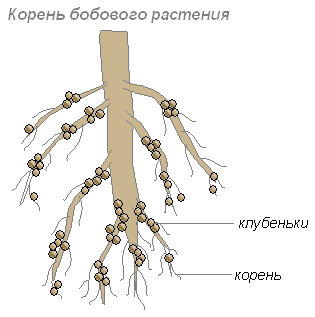
The roots provide carbohydrates to the bacteria, and the bacteria to the roots provide nitrogen-containing substances that can be absorbed by the plant. Their cohabitation is mutually beneficial.
Plant roots secrete a lot of organic substances (sugars, amino acids and others) that bacteria feed on. Therefore, especially many bacteria settle in the soil layer surrounding the roots. These bacteria convert dead plant debris into plant-available substances. This layer of soil is called the rhizosphere.
There are several hypotheses about the penetration of nodule bacteria into root tissue:
- through damage to epidermal and cortex tissue;
- through root hairs;
- only through the young cell membrane;
- thanks to companion bacteria producing pectinolytic enzymes;
- due to stimulation of the synthesis of B-indoleacetic acid from tryptophan, always present in plant root secretions.
The process of introduction of nodule bacteria into root tissue consists of two phases:
- infection of root hairs;
- process of nodule formation.
In most cases, the invading cell actively multiplies, forms so-called infection threads and, in the form of such threads, moves into the plant tissue. Nodule bacteria emerging from the infection thread continue to multiply in the host tissue.

Plant cells filled with rapidly multiplying cells of nodule bacteria begin to rapidly divide. The connection of a young nodule with the root of a legume plant is carried out thanks to vascular-fibrous bundles. During the period of functioning, the nodules are usually dense. By the time optimal activity occurs, the nodules acquire a pink color (thanks to the leghemoglobin pigment). Only those bacteria that contain leghemoglobin are capable of fixing nitrogen.
Nodule bacteria create tens and hundreds of kilograms of nitrogen fertilizer per hectare of soil.
Metabolism
Bacteria differ from each other in their metabolism. In some it occurs with the participation of oxygen, in others - without it.
Most bacteria feed on ready-made organic substances. Only a few of them (blue-green, or cyanobacteria) are capable of creating organic substances from inorganic ones. They played an important role in the accumulation of oxygen in the Earth's atmosphere.
Bacteria absorb substances from the outside, tear their molecules into pieces, assemble their shell from these parts and replenish their contents (this is how they grow), and throw unnecessary molecules out. The shell and membrane of the bacterium allows it to absorb only the necessary substances.
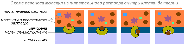
If the shell and membrane of a bacterium were completely impermeable, no substances would enter the cell. If they were permeable to all substances, the contents of the cell would mix with the medium - the solution in which the bacterium lives. To survive, bacteria need a shell that allows necessary substances to pass through, but not unnecessary substances.
The bacterium absorbs nutrients located near it. What happens next? If it can move independently (by moving a flagellum or pushing mucus back), then it moves until it finds the necessary substances.
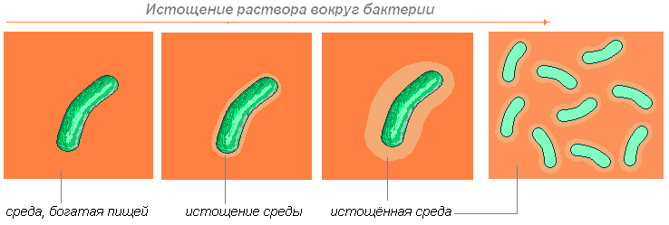
If it cannot move, then it waits until diffusion (the ability of molecules of one substance to penetrate into the thicket of molecules of another substance) brings the necessary molecules to it.
Bacteria, together with other groups of microorganisms, perform enormous chemical work. By converting various compounds, they receive the energy and nutrients necessary for their life. Metabolic processes, methods of obtaining energy and the need for materials for building the substances of their bodies are diverse in bacteria.
Other bacteria satisfy all their needs for carbon necessary for the synthesis of organic substances in the body at the expense of inorganic compounds. They are called autotrophs. Autotrophic bacteria are capable of synthesizing organic substances from inorganic ones. Among them are:
Chemosynthesis
The use of radiant energy is the most important, but not the only way to create organic matter from carbon dioxide and water. Bacteria are known that use not sunlight as an energy source for such synthesis, but the energy of chemical bonds occurring in the cells of organisms during the oxidation of certain inorganic compounds - hydrogen sulfide, sulfur, ammonia, hydrogen, nitric acid, ferrous compounds of iron and manganese. They use the organic matter formed using this chemical energy to build the cells of their body. Therefore, this process is called chemosynthesis.
The most important group of chemosynthetic microorganisms are nitrifying bacteria. These bacteria live in the soil and oxidize ammonia formed during the decay of organic residues to nitric acid. The latter reacts with mineral compounds of the soil, turning into salts of nitric acid. This process takes place in two phases.
Iron bacteria convert ferrous iron into oxide iron. The resulting iron hydroxide settles and forms the so-called bog iron ore.
Some microorganisms exist due to the oxidation of molecular hydrogen, thereby providing an autotrophic method of nutrition.
A characteristic feature of hydrogen bacteria is the ability to switch to a heterotrophic lifestyle when provided with organic compounds and the absence of hydrogen.
Thus, chemoautotrophs are typical autotrophs, since they independently synthesize the necessary organic compounds from inorganic substances, and do not take them ready-made from other organisms, like heterotrophs. Chemoautotrophic bacteria differ from phototrophic plants in their complete independence from light as an energy source.
Bacterial photosynthesis
Some pigment-containing sulfur bacteria (purple, green), containing specific pigments - bacteriochlorophylls, are able to absorb solar energy, with the help of which hydrogen sulfide in their bodies is broken down and releases hydrogen atoms to restore the corresponding compounds. This process has much in common with photosynthesis and differs only in that in purple and green bacteria the hydrogen donor is hydrogen sulfide (occasionally carboxylic acids), and in green plants it is water. In both of them, the separation and transfer of hydrogen is carried out due to the energy of absorbed solar rays.
This bacterial photosynthesis, which occurs without the release of oxygen, is called photoreduction. Photoreduction of carbon dioxide is associated with the transfer of hydrogen not from water, but from hydrogen sulfide:
6СО 2 +12Н 2 S+hv → С6Н 12 О 6 +12S=6Н 2 О
The biological significance of chemosynthesis and bacterial photosynthesis on a planetary scale is relatively small. Only chemosynthetic bacteria play a significant role in the process of sulfur cycling in nature. Absorbed by green plants in the form of sulfuric acid salts, sulfur is reduced and becomes part of protein molecules. Further, when dead plant and animal remains are destroyed by putrefactive bacteria, sulfur is released in the form of hydrogen sulfide, which is oxidized by sulfur bacteria to free sulfur (or sulfuric acid), forming sulfites in the soil that are accessible to plants. Chemo- and photoautotrophic bacteria are essential in the nitrogen and sulfur cycle.
Sporulation
Spores form inside the bacterial cell. During the process of sporulation, the bacterial cell undergoes a number of biochemical processes. The amount of free water in it decreases and enzymatic activity decreases. This ensures the resistance of the spores to unfavorable environmental conditions (high temperature, high salt concentration, drying, etc.). Sporulation is characteristic of only a small group of bacteria.

Spores are an optional stage in the life cycle of bacteria. Sporulation begins only with a lack of nutrients or accumulation of metabolic products. Bacteria in the form of spores can remain dormant for a long time. Bacterial spores can withstand prolonged boiling and very long freezing. When favorable conditions occur, the spore germinates and becomes viable. Bacterial spores are an adaptation to survive in unfavorable conditions.
Reproduction
Bacteria reproduce by dividing one cell into two. Having reached a certain size, the bacterium divides into two identical bacteria. Then each of them begins to feed, grows, divides, and so on.

After cell elongation, a transverse septum gradually forms, and then the daughter cells separate; In many bacteria, under certain conditions, after dividing, cells remain connected in characteristic groups. In this case, depending on the direction of the division plane and the number of divisions, different shapes arise. Reproduction by budding occurs as an exception in bacteria.
Under favorable conditions, cell division in many bacteria occurs every 20-30 minutes. With such rapid reproduction, the offspring of one bacterium in 5 days can form a mass that can fill all seas and oceans. A simple calculation shows that 72 generations (720,000,000,000,000,000,000 cells) can be formed per day. If converted into weight - 4720 tons. However, this does not happen in nature, since most bacteria quickly die under the influence of sunlight, drying, lack of food, heating to 65-100ºC, as a result of struggle between species, etc.
The bacterium (1), having absorbed enough food, increases in size (2) and begins to prepare for reproduction (cell division). Its DNA (in a bacterium the DNA molecule is closed in a ring) doubles (the bacterium produces a copy of this molecule). Both DNA molecules (3,4) find themselves attached to the wall of the bacterium and, as the bacterium elongates, move apart (5,6). First the nucleotide divides, then the cytoplasm.

After the divergence of two DNA molecules, a constriction appears on the bacterium, which gradually divides the body of the bacterium into two parts, each of which contains a DNA molecule (7).
It happens (in Bacillus subtilis) that two bacteria stick together and a bridge is formed between them (1,2).

The jumper transports DNA from one bacterium to another (3). Once in one bacterium, DNA molecules intertwine, stick together in some places (4), and then exchange sections (5).
The role of bacteria in nature
Gyre
Bacteria are the most important link in the general cycle of substances in nature. Plants create complex organic substances from carbon dioxide, water and mineral salts in the soil. These substances return to the soil with dead fungi, plants and animal corpses. Bacteria break down complex substances into simple ones, which are then used by plants.
Bacteria destroy complex organic substances of dead plants and animal corpses, excretions of living organisms and various wastes. Feeding on these organic substances, saprophytic bacteria of decay turn them into humus. These are a kind of orderlies of our planet. Thus, bacteria actively participate in the cycle of substances in nature.
Soil formation
Since bacteria are distributed almost everywhere and occur in huge numbers, they largely determine various processes occurring in nature. In autumn, the leaves of trees and shrubs fall, above-ground shoots of grasses die, old branches fall off, and from time to time the trunks of old trees fall. All this gradually turns into humus. In 1 cm3. The surface layer of forest soil contains hundreds of millions of saprophytic soil bacteria of several species. These bacteria convert humus into various minerals that can be absorbed from the soil by plant roots.
Some soil bacteria are able to absorb nitrogen from the air, using it in vital processes. These nitrogen-fixing bacteria live independently or settle in the roots of legume plants. Having penetrated the roots of legumes, these bacteria cause the growth of root cells and the formation of nodules on them.
These bacteria produce nitrogen compounds that plants use. Bacteria obtain carbohydrates and mineral salts from plants. Thus, there is a close relationship between the legume plant and the nodule bacteria, which is beneficial to both one and the other organism. This phenomenon is called symbiosis.
Thanks to symbiosis with nodule bacteria, leguminous plants enrich the soil with nitrogen, helping to increase yield.
Distribution in nature
Microorganisms are ubiquitous. The only exceptions are the craters of active volcanoes and small areas at the epicenters of exploded atomic bombs. Neither the low temperatures of Antarctica, nor the boiling streams of geysers, nor saturated salt solutions in salt pools, nor the strong insolation of mountain peaks, nor the harsh irradiation of nuclear reactors interfere with the existence and development of microflora. All living beings constantly interact with microorganisms, often being not only their repositories, but also their distributors. Microorganisms are natives of our planet, actively exploring the most incredible natural substrates.
Soil microflora
The number of bacteria in the soil is extremely large - hundreds of millions and billions of individuals per gram. There are much more of them in soil than in water and air. The total number of bacteria in soils changes. The number of bacteria depends on the type of soil, their condition, and the depth of the layers.
On the surface of soil particles, microorganisms are located in small microcolonies (20-100 cells each). They often develop in the thickness of clots of organic matter, on living and dying plant roots, in thin capillaries and inside lumps.
The soil microflora is very diverse. Here there are different physiological groups of bacteria: putrefaction bacteria, nitrifying bacteria, nitrogen-fixing bacteria, sulfur bacteria, etc. among them there are aerobes and anaerobes, spore and non-spore forms. Microflora is one of the factors in soil formation.
The area of development of microorganisms in the soil is the zone adjacent to the roots of living plants. It is called the rhizosphere, and the totality of microorganisms contained in it is called the rhizosphere microflora.
Microflora of reservoirs
Water is a natural environment where microorganisms develop in large numbers. The bulk of them enters the water from the soil. A factor that determines the number of bacteria in water and the presence of nutrients in it. The cleanest waters are from artesian wells and springs. Open reservoirs and rivers are very rich in bacteria. The largest number of bacteria is found in the surface layers of water, closer to the shore. As you move away from the shore and increase in depth, the number of bacteria decreases.
Clean water contains 100-200 bacteria per ml, and polluted water contains 100-300 thousand or more. There are many bacteria in the bottom sludge, especially in the surface layer, where the bacteria form a film. This film contains a lot of sulfur and iron bacteria, which oxidize hydrogen sulfide to sulfuric acid and thereby prevent fish from dying. There are more spore-bearing forms in silt, while non-spore-bearing forms predominate in water.
In terms of species composition, the microflora of water is similar to the microflora of soil, but there are also specific forms. By destroying various waste that gets into the water, microorganisms gradually carry out the so-called biological purification of water.
Air microflora
The microflora of the air is less numerous than the microflora of soil and water. Bacteria rise into the air with dust, can remain there for some time, and then settle on the surface of the earth and die from lack of nutrition or under the influence of ultraviolet rays. The number of microorganisms in the air depends on the geographical zone, terrain, time of year, dust pollution, etc. each speck of dust is a carrier of microorganisms. Most bacteria are in the air above industrial enterprises. The air in rural areas is cleaner. The cleanest air is over forests, mountains, and snowy areas. The upper layers of air contain fewer microbes. The air microflora contains many pigmented and spore-bearing bacteria, which are more resistant than others to ultraviolet rays.
Microflora of the human body
The human body, even a completely healthy one, is always a carrier of microflora. When the human body comes into contact with air and soil, various microorganisms, including pathogenic ones (tetanus bacilli, gas gangrene, etc.), settle on clothing and skin. The most frequently exposed parts of the human body are contaminated. E. coli and staphylococci are found on the hands. There are over 100 types of microbes in the oral cavity. The mouth, with its temperature, humidity, and nutrient residues, is an excellent environment for the development of microorganisms.
The stomach has an acidic reaction, so the majority of microorganisms in it die. Starting from the small intestine, the reaction becomes alkaline, i.e. favorable for microbes. The microflora in the large intestines is very diverse. Each adult excretes about 18 billion bacteria daily in excrement, i.e. more individuals than people on the globe.
Internal organs that are not connected to the external environment (brain, heart, liver, bladder, etc.) are usually free of microbes. Microbes enter these organs only during illness.
Bacteria in the cycle of substances
Microorganisms in general and bacteria in particular play a large role in the biologically important cycles of substances on Earth, carrying out chemical transformations that are completely inaccessible to either plants or animals. Different stages of the cycle of elements are carried out by organisms of different types. The existence of each individual group of organisms depends on the chemical transformation of elements carried out by other groups.
Nitrogen cycle
The cyclic transformation of nitrogenous compounds plays a primary role in supplying the necessary forms of nitrogen to organisms of the biosphere with different nutritional needs. Over 90% of total nitrogen fixation is due to the metabolic activity of certain bacteria.
Carbon cycle
The biological transformation of organic carbon into carbon dioxide, accompanied by the reduction of molecular oxygen, requires the joint metabolic activity of various microorganisms. Many aerobic bacteria carry out complete oxidation of organic substances. Under aerobic conditions, organic compounds are initially broken down by fermentation, and the organic end products of fermentation are further oxidized by anaerobic respiration if inorganic hydrogen acceptors (nitrate, sulfate, or CO 2 ) are present.
Sulfur cycle
Sulfur is available to living organisms mainly in the form of soluble sulfates or reduced organic sulfur compounds.
Iron cycle
Some freshwater bodies contain high concentrations of reduced iron salts. In such places, a specific bacterial microflora develops - iron bacteria, which oxidize reduced iron. They participate in the formation of bog iron ores and water sources rich in iron salts.
Bacteria are the most ancient organisms, appearing about 3.5 billion years ago in the Archean. For about 2.5 billion years they dominated the Earth, forming the biosphere, and participated in the formation of the oxygen atmosphere.
Bacteria are one of the most simply structured living organisms (except viruses). They are believed to be the first organisms to appear on Earth.
According to the new system, three domains are distinguished: “Bacteria” (eubacteria), “Archaea” (archaebacteria) and “Eisagua” (eukaryotes). Domains include phyla, classes, orders, families, genera, and species.
Bacteria are prokaryotes (in a prokaryotic cell, the nucleus, called the nucleoid, does not have a nuclear envelope, nucleolus and histones, and the cytoplasm does not contain highly organized organelles).
To thick-walled, gram-positive bacteria include: spherical forms, or cocci (staphylococci, streptococci, pneumococci); rod-shaped, including corynebacteria, mycobacteria and bifidobacteria; actinomycetes (branching, filamentous bacteria).
Spherical forms, or cocci - spherical bacteria 0.5-1.0 microns in size; According to the relative arrangement of cells, micrococci, diplococci, streptococci, tetracocci, sarcina and staphylococci. Micrococci (Greek) mikros- small)
- separately located cells or in the form of “packages”.
Diplococci (from Greek.diploos- double), or paired cocci, arranged in pairs (pneumococcus, gonococcus, meningococcus), since the cells do not diverge after division. Pneumococcus has a lanceolate shape on opposite sides, and gonococcus And meningococcus have the shape of coffee beans with their concave surface facing each other. Streptococci(from Greek streptos- chain) - cells of a round or elongated shape, forming a chain due to cell division in the same plane and maintaining the connection between them at the site of division. Sarcins (from lat. sarcina- bunch, bale) are arranged in the form of “packets” of 8 or more cocci, since they are formed during cell division in three mutually perpendicular planes. Staphylococcus (from Greek. staphyle- grape bunch) - cocci located in the form of a bunch of grapes as a result of division in different planes.
Rod-shaped bacteria differ in size, shape of cell ends and relative position of cells. The length of the cells varies from 1.0 to 8.0 µm, thickness - from 0.5 to 2.0 µm. Rods can be regular (Escherichia coli, etc.) and irregular (corynebacteria, etc.) shapes, including branching ones, for example in actinomycetes. The slightly curved rods are called vibrios (Vibrio cholerae). Most rod-shaped bacteria is located randomly, since after division the cells diverge. If, after division, the cells remain connected by common fragments of the cell wall and do not diverge, then they are located at an angle to each other (Corynebacterium diphtheria) or form a chain (anthrax bacillus).
Rice. 3.1. SmearfromEscherichia coliAndStaphylococcus aureus.Gram painting
In the old Bergey Manual of Systematic Bacteriology, bacteria were divided according to the characteristics of the bacterial cell wall into 4 divisions: Gracilicutes - eubacteria with a thin cell wall, gram-negative; Firmicutes - eubacteria with a thick cell wall, gram-positive; Tenericutes - eubacteria without a cell wall; Mendosicutes are archaebacteria with a defective cell wall. Each department was divided into sections, or groups, based on Gram staining, cell shape, oxygen demand, motility, metabolic and nutritional characteristics.
Table 3.1. According to the 2nd edition (2001) of the Bergey Manual, bacteria are divided into two domains: “Bacteria»
And «
Archaea»
|
Domain "Bacteria"(eubacteria) |
Domain "Archaea"(archaebacteria) |
|
In the domain «
Bacteria»
The following bacteria can be isolated: |
Archaebacteria do not contain peptidoglycan in their cell wall. They have special ribosomes and ribosomal RNA (rRNA). The term “archaebacteria” appeared in 1977. This is one of the ancient forms of life, which is what the prefix “arche” means. There are no pathogens of infectious diseases among them. |
|
‘ Most Gram-negative bacteria are grouped into the phylum Proteobacteria, based on similarities in ribosomal RNA (“Proteobacteria” - from the Greek god Proteus, who took various forms). They evolved from a common photosynthetic ancestor. |
|
To domain "Bacteria"includes 22 types, of which the following are of medical importance (according to Bergi, 2001):
TypeINIV. Deinococcus-ThermusClassI. Deinococci
Order I. Deinococcales Family I. Deinococcaceae Genus1. Deinococcus
TypeINXII. Proteobacteria
ClassI. Alphaproteobacteria Order II. Rickettsiales Family I. Rickettsiaceae Genus I. Rickettsia Genus I. Orientia Genus III. Wolbachia Family I. Ehrlichiaceae (b genera) Genus I. Ehrlichia Genus I. Aegyptianella Family III. Holosporaceae (8 genera) Order VI. Rhizobiales Family I. Bartonellaceae Genus I. Bartonella Family III. Brucellaceae Genus I. Brucella
ClassII. Betaproteobacteria Order I. Burkholderiales Family I. Burkholderiaceae Genus I. Burkholderia Family IV. Alcaligenaceae Genus I. Alcaligenes Genus III. Bordetella Order IV. Neisseriales Family I. Neisseriaceae Genus I. Neisseria Genus VI.Eikenella Genus IX.Kingella Order V. Nitrozomonadales Family II. Spirillaceae Genus I. Spirillum
ClassIII. Gammaproteobacteria Order V. Thiotrichales Family III. Francisellaceae Genus I. Francisella Order VI. Legionellales Family I. Legionellaceae Genus I. Legionella Family II. Coxiellaceae Genus I. Coxiella Order IX. Pseudomonadales Family I. Pseudomonadaceae Genus I. Pseudomonas Family I. Moraxellaceae Genus I. Moraxella Genus I. Acinetobacter Order XI. Vibrionales Family I. Vibrionaceae Genus I. Vibrio Order XII. Aeromonadales Family I. Aeromonadaceae Genus I. Aeromonas Order XIII. Enterobacteriales Family I. Enterobacteriaceae Genus I. Enterobacter Genus VIII. Calymmatobacterium Genus X. Citrobacter Genus XI. Edwardsiella Genus XII. Erwinia Genus XIII Escherichia Genus XV. Hafnia
Genus XVI. Klebsiella Genus XVII. Kluyvera Genus XXI. Morganella Genus XXVI. Plesiomonas Genus XXVIII. Proteus Genus XXIX. Providencia RodXXXY. Salmonella Genus XXXIII. Serratia Genus XXXIV. Shigella Genus XL. Yersinia Order IV. Pasteurellales Family I. Pasteurellaceae Genus I. Pasteurella Genus I. Actinobacillus Genus III. Haemophilus
ClassIV. Deltaproteobacteria Order II. Desulfovibrionales Family I. Desulfovibrionaceae Genus II. Bilophila
ClassV. Epsilonproteobacteria Order I. Campylobacteriales Family I. Campylobacteriaceae Genus I. Campylobacter Family II. Helicobacteriaceae Genus I. Helicobacter Genus Il.Wolinella
TypeINXIII. Firmicutes (mainwaygram-positive)
ClassI.
Clostridia Order I. Clostridiales Family I. Clostridiaiaceae Genus I. Clostridium Genus IX. Sarcina Family III. Peptostreptococcaceae Genus I. Peptostreptococcus Family IV. Eubacteriaceae Genus I. Eubacterium Family V. Peptococcaceae Genus I. Peptococcus Family VII. Acidaminococcaceae Genus XIV. Veillonella ClassII. Mollicutes Order I. Mycoplasmatales Family I. Mycoplasmataceae Genus I. Mycoplasma Genus IV. Ureaplasma ClassIII. Bacilli Order I. Bacilalles Family I. Bacillaceae Genus I. Bacillus Family I. Planococcaceae Genus I. Planococcus Genus IV. Sporosarcina Family IV. Listeriaceae Genus I. Listeria Family V. Staphylococcaceae Genus I. Staphylococcus Genus II. Gemella Order I. Lactobacillales Family I. Lactobacillaceae Genus I. Lactobacillus Genus III. Pediococcus Family II. Aegococcusae Genus I. Aerococcus Family IV. Enterococcaceae Genus I. Enterococcus
Family V. Leuconostocaceae Genus I. Leuconostoc Family VI. Streptococcaceae Genus I. Streptococcus Genus II. Lactococcus TypeINXIV. ActinobacteriaClassI. Actinobacteria Subclass V. Actinobacteridae Order I. Actinomycetales Suborder V. Actinomycineae Family I. Actinomycetaceae Genus I. Actinomyces Genus I. Actinobacilum Genus III. Arcanodacterium Genus IV. Mobiluncus Suborder VI. Micrococcineae Family I. Micrococcaceae Genus I. Micrococcus Genus VI. Rothia Genus VII. Stomatococcus Suborder VII. Corynebacterineae Family I. Corynebacteriaceae Genus I. Corynebacterium Family I. Mycobacteriaceae Genus IV. Mycobacterium Family V. Nocardiaceae Genus I. Nocardia Genus I. Rhodococcus Suborder VII. Propionibacterineae Family I. Propionibacteriaceae Genus I. Propionibacterium Family I. Nocardiaceae Genus I. Nocardioides Order II. Bifidobacteriales
Family I. Bifidobacteriaceae Genus I. Bifidobacterium Genus III. Gardnerella TypeINXVI. ChlamydiaeClassI. Chlamydiae Order I. Chlamydiales Family I. Chlamydiaceae Genus I. Chlamydia Genus II. Chlamydophila TypeINXVII. SpirochaetesClassI. Spirochaetes Order I. Spirochaetales Family I. Spirochaetaceae Genus I. Spirochaeta Genus I. Borrelia Genus IX. Treponema Family III. Leptospiraceae Genus I. Leptospira TypeINXX. BacteroidetesClassI. Bacteroidetes Order I. Bacteroidales Family I. Bacteroidaceae Genus I. Bacteroides Family III. Porphyromonadaceae Genus I. Porphyromonas Family IV. Prevotellaceae Genus I. Prevotella ClassII. Flavobacteria Order I. Flavobacteriales Family I. Flavobacteriaceae Genus I. Flavobacterium

*Spore location: 1 - central, 2 - subterminal, 3 - terminal.
Rice. 3.2.
Chlamydia belong to obligate intracellular coccoid gram-negative (sometimes gram-variable) bacteria. They reproduce only in living cells. Outside the cells, chlamydia are spherical in shape (0.3 µm), metabolically inactive and are called elementary bodies. The cell wall of elementary bodies contains a major outer membrane protein and a protein containing a large amount of cysteine. Elementary bodies enter the epithelial cell by endocytosis with the formation of an intracellular vacuole. Inside the cells, they enlarge and transform into dividing reticular bodies, forming clusters in vacuoles (inclusions). Elementary bodies are formed from reticular bodies, which leave the cells by exocytosis or cell lysis. Elementary bodies released from the cell enter a new cycle, infecting other cells. In humans, chlamydia causes damage to the eyes, urogenital tract, lungs, etc.
Mycoplasma - small bacteria (0.15-1.0 µm), surrounded by a cytoplasmic membrane and without a cell wall. Due to the absence of a cell wall, mycoplasmas are osmotically sensitive. They have a variety of shapes: coccoid, filamentous, flask-shaped; similar to L-shapes (Fig. 3.6). These forms are visible under phase-contrast microscopy of pure cultures of mycoplasmas. Pathogenic mycoplasmas cause chronic infections - mycoplasmosis.
Actinomycetes - branching, filamentous or rod-shaped gram-positive bacteria. Its name (from Greek. actis- Ray, mykes- fungus) they received due to the formation in the affected tissues of drusen - granules of tightly intertwined threads in the form of rays extending from the center and ending in flask-shaped thickenings. Actinomycetes can divide by fragmentation of the mycelium into cells similar to rod-shaped and coccoid bacteria. On the aerial hyphae of actinomycetes, spores can form that serve for reproduction. Actinomycete spores are usually not heat-resistant.

Attachment site (analogous to blepharoplast)
Rice. 3.3.Electron diffraction pattern of a Treponema pallidum cell fragment (negative contrast). According to N. M. Ovchinnikov, V. V. Delectorsky
A common phylogenetic branch with actinomycetes is formed by the so-called nocardiform (nocardioform) actinomycetes - a collective group of rod-shaped, irregularly shaped bacteria. Their individual representatives form branching forms. These include bacteria of the genera Corynebacterium,
Mycobacterium,
Nocardia etc. Nocardi-like actinomycetes are distinguished by the presence in the cell wall of the sugars arabinose, galactose, as well as mycolic acids and large amounts of fatty acids. Mycolic acids and cell wall lipids determine the acid resistance of bacteria, in particular, mycobacteria tuberculosis and leprosy (when stained according to Ziehl-Neelsen, they are red, and non-acid-resistant bacteria and tissue elements, sputum are blue).
Convoluted forms are spiral-shaped bacteria, for example spirilla, which have the appearance of corkscrew-shaped convoluted cells. Pathogenic spirilla includes the causative agent sodoku (rat bite disease). The convoluted ones also include Campylobacter, Helicobacter, which have curves like the wing of a flying seagull; bacteria such as spirochetes are also close to them. Spirochetes are thin, long, convoluted (spiral-shaped) bacteria that differ from spirillum in their mobility due to flexural changes in cells. Spirochetes have an outer membrane cell wall surrounding a protoplasmic cylinder with a cytoplasmic membrane. Under the outer membrane of the cell wall (in the periplasm) there are periplasmic fibrils (flagella), which, as if twisting around the protoplasmic cylinder of the spirochete, give it a helical shape (primary curls of the spirochete). The fibrils are attached to the ends of the cell (Fig. 3.3) and are directed towards each other. The other end of the fibrils is free. The number and arrangement of fibrils varies among species. Fibrils are involved in the movement of spirochetes, giving the cells rotational, bending and translational motion. In this case, spirochetes form loops, curls, and bends, which are called secondary curls.
Spirochetes do not accept dyes well. They are stained using the Romanowsky-Giemsa method or silver plating, and examined in their living form using phase-contrast or dark-field microscopy. Spirochetes are represented by 3 genera that are pathogenic for humans: Treponema, Borrelia, Leptospira.
Treponema(genus Treponema) have the appearance of thin, corkscrew-twisted threads with 8-12 uniform small curls. Fibrils are located around the protoplast of the treponema. Pathogenic representatives are T. pallidum- the causative agent of syphilis, T. pertenue- causative agent of the tropical disease yaws.
Borrelia(genus Borrelia) are longer, have 3-8 large whorls and 8-20 fibrils. These include the causative agent of relapsing fever (B. recurrentis) and pathogens of Lyme disease (V. burgdorferi and etc.).
Leptospira(genus Leptospira) have shallow and frequent curls - in the form of a twisted rope. The ends of these spirochetes are curved like hooks with thickenings at the ends. Forming secondary curls, they take on the appearance of the letters “S” or “C”; have 2 axial threads. Pathogenic representative L
