Vertebral pole.
The spine (Columna Vertebralis) is formed from 31-32 vertebrae (Vertebrae). Vertebrae Thoracicae (VerteBrae Lumbales), 5 Sacralas (VerteBrae Sacrales), 5 Sacralas, Sacrum, and 2 - 3 Copshings (VerteBrae Coccygeae) distinguish vertebra
Vertebrae
35. Chest vertebra (VIII).
1 - Processus ARTICULARIS SUPERIOR;
2 - FOVEA COSTALIS SUPERIOR;
3 - Corpus Vertebrae;
4 - FOVEA COSTALIS INFERIOR;
5 - Incisura Vertebralis Interior;
6 - Processus ARTICULARIS INFERIOR;
7 - Processus Spinosus;
8 - Processus Transversus;
9 - FOVEA COSTALIS TRANSVERSALIS.
Breast vertebrae (Vertebrae Thoracicae) (Fig. 35). They are tested with rear ends of the ribs. They differ from the lumbar vertebrae by the fact that the transverse dimensions of their bodies are less. The shape of the bodies of breast vertebrae is approaching a triangle. At the top and bottom edges of the side parts of the body lie (FOVEA COSTALIS SUPERIOR ET INFERIOR). Top and lower pits - places for articulation with the head of the corresponding rib. In I, the vertebra has a fossa on the upper edge for compounding with the I edge and in the lower edge - to connect with the second edge. X The vertebra has a hole only in the upper edge. XI and XII breast vertebrae have one hole for appropriate ribs. A arc (Arcus Vertebrae) with two legs (Pedunculi Arcus Vertebrae), having small cuts, is attached to the back surface of the body of the vertebra. The arc limits the rear vertebral hole (for. Vertebrele). From the arc to the right and left the transverse procesversi (Processus Transversi). They are well developed, which is explained by a more significant load in connection with the attachment of edges to them. On the front side of the I - X transverse processes closer to their top there are on the articular pit (FOVEA COSTALIS TRANSVERSALIS) - the place of articulation with the ribs. Back directed by a spinous process (Processus Spinosus). It begins on the back surface of the arc, turned back and down, thinner and already than the corresponding reproduction of the lumbar vertebra. From the upper and lower edges of the arc paired top and bottom articulates (Processus Articulares Superiores et Inferiores). The articular areas are located in the frontal plane.

36. Lumbar vertebra (III).
1 - Corpus Vertebrae;
2 - Incisura Vertebralis Interior;
3 - Processus ARTICULARIS INFERIOR;
4 - Processus Spinosus;
5 - Processus Costarius;
6 - Processus ARTICULARIS SUPERIOR;
7 - Incisura Vertebralis Superior.
Lumbar vertebrae (Vertebrae Lumbales) (Fig. 36). The lumbar vertebra has the greatest sizes of the body and the sausage process.
The body (Corpus) of the oval shape, its width prevails over the height. Arc (Arcus Vertebrae) is attached to the back of its surface (Pedunculi Arcus Vertebrae), which are involved in the formation of the spine (for. Vertebrale) having an oval or rounded form. The process of the vertebrae is attached to the process: rear - spinosi (Processus spinosi), having a wide plate flattened from sides, and somewhat thickened at the end, on the right and left - transverse processessus transversi, top and bottom - paired joints (Processus Articulares) . In III - V vertebrae, the articular surfaces of the processes have an oval form.
Attaching the arc legs to the body of the vertebra there are cuttings, the lower edge is more noticeable than on the top (Incisura Vertebralis Superior et Inferior), which in general the spinal column limit the intervertebral hole (for. Intervertebrile).

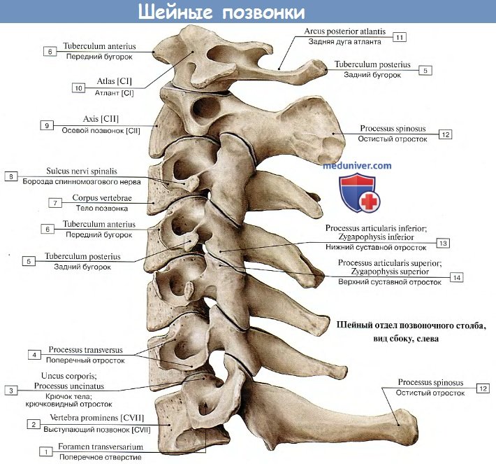
37. Cervical vertebra (VI).
1 - Corpus Vertebrae;
2 - Tuberculum Anterius;
3 - Tuberculum Posterius;
4 - Processus Spinosus;
5 - Processus ARTICULARIS SUPERIOR.
Cervical vertebrae (Vertebrae Cervicales). I and II cervical vertebrae have specific traits Buildings are described independently. III - VII The cervical vertebrae (Fig. 37) is reminded by the structure of the structure of the thoracic and lumbar vertebrae, differing from the last dimensions of the parts. The top edge of the body of the cervical vertebrae is groomed in the sagittal plane, the transverse processes are presented in the form of anterior tuberculum (reduced ribs), rear tuberculum (rearculum posterius) (reduced transverse processes), and between them there is a transverse hole (for. Transversum) . The tops of the ostic processes are twisted. At the VII vertebra, a fishing process is more than the process of other vertebrae, and palpacles through the skin, so VII vertebrae received the name of the speaker (Vertebra Prominens).

38. Cervical vertebra (I).
1 - Arcus Anterior;
2 - FOVEA ARTICULARIS INFERIOR;
3 - for. TRANSVERSARIUM;
4 - Processus Transversus;
5 - Arcus Posterior;
6 - Processus Costarius;
7 - FOVEA DENTIS.
The first cervical vertebra is Atlant (fig. 38) has anterior and rear arc (Arcus Anterior et Posterior), which are combined with paired lateral masses (Massae Laterales). On the upper and lower surfaces of the side thickens are made of articular areas: the upper ellipsoid form is the place of articulation with the tipping mysteries, the lower spherical-is the place of connection with the articular surface of the cervical vertebra. The body of the I vertebra is absent. On the front arc, there is an front tuberculum (tuberculum anterius), on the back surface of the arc - a tooth pamper (FOVEA DENTIS), a place of articulation with a dental process II vertebra. On the back arc there is a rear tuberculum (Tuberculum Posterius).

39. Cervical vertebra (II).
1 - Corpus Vertebrae;
2 - Fades Articularis Anterior;
3 - DENS;
4 - Fades Articularis Posterior;
5 - Lamina Arcus Vertebrae;
6 - Processus Spinosus;
7 - Processus Articularis Inferior;
8 - Processus Transversus;
9 - for. TRANSVERSARIUM;
10 - Fades Articularis Superior)
Second cervical vertebra - axial vertebra (AXIS) (Fig. 39).
On the upper surface of his body there is a dental extension (DENS), which is the body of the first cervical vertebra moving here. Outside and rear on the tooth there are two, front and rear, articular surfaces (Fades Articulares Anterior et Posterior) for the formation of joints with a hole of the anterior arc of Atlanta and its transverse ligament (Lig. Transversum).
Sacrum (SACRUM) (Fig. 40) After 16 years, 19 vertebrae of the sacrum of the spine has grown. Its upper part is expanded, articular processes and entrance to the sacral canal are visible. The lower part of the sacrum is narrowed, it has a hole of the sacrilate channel. On the front concave and rear convex surfaces of the sacrum there are 4 pairs of holes (Forr. Sacralia Pelvina et Dorsalia) similar to the intervertebral holes. The bone substance located the lateral of these holes (Massae Laterales) is formed by the battle of rudiment ribs and transverse vertebrae processes. On the side surfaces of the sacrum are the articular fields of the seedless shape (Facies Auriculares) are located behind them (tuberositas sacrales). On the back surface of the sacrum from the combat of splicing processes, the median sacral comb (Crista Sacralis Mediana) is formed, the articular is an intermediate sacral comb (Crista Sacralis Intermedia), a cross-lateral sacral comb (Crista Sacralis Lateralis).

40. Cresan. A - Front view: 1 - Basis Ossis SACRI; 2 - Processus ARTICULARIS SUPERIOR; 3 - Pars Lateralis; 4 - Lineae Transversae; 5 - FORR. Sacralia Pelvina; 6 - Apex Ossis Sacri. B - Rear view: 1 -Canalis Sacralis; 2 - Processus ARTICULARIS SUPERIOR; 3 - Tuberositas Sacralis; 4 - Crista Sacralis Intermedia; 5 - Crista Sacralis Mediana; 6 - HIATUS SACRALIS; 7 - Cornu Sacrale; 8 - Forr. Sacralia Dorsalia; 9 - Crista Sacralis Lateralis.
Coccyx (OS CoccyGis) is formed by incomprehension 2-3 vertebrae and is connected to the tip of the sacrum.
Ossification. From the ventromedal surface of somites (see the initial stages of embryogenesis), a group of mesenchymal cells, which surround chord, giving a derivative of the vertebrae, is combined in the sclerot. Of the two adventures, the cartilaginous core of the body of the future vertebra is formed at the place of their contact of their contact. Such secondary segmentation contributes to the fact that the Miotoma will grow with their ends with two next to the lying aliases (Fig. 41). At the 6th week of embryonic development on the site of the mesenchymal laying, a cartilage fabric is formed. The first cores of ossification appear in the body of the XII of the breast vertebra at the 6-7th week. In the rest of the chest and lumbar vertices, the osenation core arise by the end of the 12th week, in the cervical and two top sacrats - at the end of the 16th week. At this time, three paired cores of ossification are formed in the cartest of the vertebral opening: the legs of the arc are formed from the front, the arc plate and the base of the spinner process, from the transverse nucleus - the base of the transverse process. Only on the 2nd year of life, starting with the cervical vertebrae, a complete bone arc is formed. The 4-year-old child is still widened arcs of the cervical, V lumbar, I, IV and V sacral vertebrae. Their closure occurs on the 7th year.

41. Scheme of the development of the vertebrae (by Clar). 1 - Somit; 2 - Miot; 3 - Discus intervertebralis; 4 - muscles; 5 - vertebrae developing from parts of two somits.

42. Scheme of the lumbar vertebra (on Andronesc).
1 - primary middle core;
2 - upper epiphyseal ring of ossification;
3 - lower epiphyseal ring;
4 - primary front-flying and transverse cores of ossification;
5 - secondary Lower Undercut;
6 - primary rear agent core;
7 - the secondary core of the ossification of the cooler process;
8 - secondary transverse core;
9 - the secondary core of the silence of the maternity process;
10 is a secondary ultimate oscillage core.
IN adolescence The bodies of the vertebrae are secondary cores of osenation, having a type of plates (epiphyseal rings) (Fig. 42). Starting from 15 years, initially in breast vertebrae and ending with lumbar occurs synostosition of epiphyseal rings to the bodies of the vertebrae.
Some feature represents the ossification of I and II cervical vertebrae. At the 16th week, two primary kernels appear in the tooth, which grow up with the body of the vertebra only on the 4th and5th year of life.
Anomalies. Most often, the emergence of the vertebral development abnormalities is the missessession of their arcs (spondylolysis) mainly in the sacrus, which contributes to the development of Spina Bifida. Less often there is an outset of halves of vertebrae bodies with each other. There is a complete absence of the bodies of the vertebrae (asomia), the absence of half the body of the vertebra (hemisomy), the cessation of the growth of the body of the vertebrae in height (congenital platinum).
In Latin, six cases:
Nominatīvus.calm Who? what?
Genetīvus.pAGE WHO? What?
Datīvus.duty to whom? What?
Accusatīvus. Request? what?
Ablatīvus. Decaying by whom? than? OK? about what?
Vocatīvus. vocative
For the correct understanding of the majority of the anatomical terms (and the terms of the other sections of medical terminology), it is enough to know only the forms of the first two cases of the sole and multiple numberWe subsequently restrictly:
The nominative case is the name of the name, names, is considered the initial form of nouns and adjectives. In anatomy histological terms, nouns in the nominal case are written in the first place.
The system change in the numbers and cases is called declining. In Latin, there are five types of changes in words by numbers and cases or five decons.
The declination of the Latin nouns is made to determine at the end of the genitive case of the only number - Gen. Sing. Since only in this case, every declination has a characteristic ending. In other cases, depending on the type and nature of the foundation, the foundations may coincide or have several options ( see Consolidated Case Study Table).
Nouns declining table
Finishing Gen.Sing. | |||||
Declining |
Ending Gen.. sing.. (the genitive case of the only number) is always recorded in the nouns in the dictionary.
Vocational form of nouns
Vocabulary form of nouns is the following entry: Costa, AE F edge; Muscŭlus, i m muscle; Sternum, I N sternum; Margo, ĭnis m edge; Arcus, US M arc; FACIES, EI F face, surface; Where the whole word recorded at the beginning is the form of a nominative case of a single number, through the occupied - the end of the genitive case of the only number, and the letter is denoted by the generation of this noun. For some nouns (more often than the 3rd decline) in the parental case is recorded not only padded endingBut also part of the foundation to designate cases when alternating vowels or consonant sounds are based on words. For example: Corpus, ŏris n body; FORāMEN, ĭNIS N hole; Apex, ĭcis m top. If only one syllable word has a word in the nominative case, the form of the genitive case is recorded in full: OS, OSSIS N bone; OS, ORIS N mouth; DENS, DENTIS M tooth; Pars, Partis F part.Therefore, when memorizing the Latin nouns, it is necessary to memorize not only the initial form, but also the form of a genitive case, and what kind of this word: Costa, Costae, femin.ī nUM.; Forāmen, ForaMĭnis, neutrum; Margo, Margĭnis, mascul.ī nUM..
NOM . sing. . | Ending Gen. . sing. | nouns | |
edge |
|||
muscle |
|||
sternum |
|||
edge |
|||
arc |
|||
face, surface |
|||
bone |
|||
part |
When memorizing the Latin nouns, it is necessary to memorize all the elements of the vocabulary form. Thus, the forms of the first two cases, which are most found in the anatomical terms, we will know only on the basis of the knowledge of the vocabulary form of the noun.
Greek nouns in anatomical nomenclature
In the anatomical terminology, the Greek nouns passed to the Latin language, which are divided into three decons. The fission is based on the same principle as Latin nouns: the end of the genius of the only number. When declining the Greek words mostly take the Latin endings, but in some cases they retain the old, Greek: ALO, ES F aloe (Medicinal Plant) ; Raphe, ES F the seam; Diabētes, ae m diabetes; ASCītes, ae m vodkyanka abdominal cavity.Such words will be considered within the framework of Latin decline.
To secure a new material:
Determine declining nouns : vERTĕBRA, AE F; Corpus, ŏris n; Dorsum, I N; Arcus, US M; Superficies, ēi F; Basis, IS F; Collum, I n; Apex, ĭcis m; CRANIUM, II N; DUCTUS, US M; Caput, ĭtis n; Ganglion, II N; CORNU, US N; Squama, AE F; Facies, ēi F; zygōma, ătis n; Processus, US M; tubercŭlum, I n; Thorax, ācis m; TRACTUS, US M; atlas, antis m; Axis, IS M; Dorsum, I N; Genu, us n.
§nine. Structure of anatomical terms.
Inconsistent definition
1) Anatomical terms may consist of one word. We will call them alone - vertěbra vertebra; Costa. edge; Cerěbrum. brainetc . You need to know that some single Latin names into Russian are transferred not by one Russian word, but two. For example: Thorax (in Greek pancir) - rib cage;fibula (Latin clotter pin to whom bone looks like) - fibula; Tibia (Latin dudge, which in antiquity made from such bones) - Tibra Tight bone, etc.
2) Twoldal terms consist of two words: Corpus Vertěbrae body (what?) vertebra; vertěbra Cervicālis vertebra (what?) cervical etc. In two-bed terms, the first word is always a noun in the nominal case - NOM. Sing. The second word will determine, characterizes the first, it is called definition. The definition expressed by nouns in the parental case is called an inconsistent definition.
3) Multile terms consist of several nouns and adjectives: Facies Articulāris Tubercŭli Costae the articular surface of the tubercle ribs. In the Latin term, the noun in the nominative case is first, although in Russian first we call adjective.
§10. Sequence of actions when transferring to Latin
terms with checked definition
Any anatomical term in Latin starts with a noun in the nominal case of a single or multiple number. Next, follow the words explaining this noun. These may be adjectives (consistent definition) or nouns in the parental case (inconsistent definition).
The simplest design is the "noun nougeny case + noun genitive padege." Denote them from 1 and C 2. And in Russian and in Latin, the words are located in the same sequence "C 1 + C 2".
Consider for example the translation of the term arc Rib .
First of all, it is necessary to recall the vocabulary form of each word included in the term:
arc - Arcus, US M;
rib - Costa, AE F
Then you need to determine which case is used in this term every word in Russian, and in the same case, write the Latin word:
Connect Latin forms according to the scheme "C 1 + C 2" and get the Latin term as a result arcus costae. .
Anatomical term may include several words in the parental case: the surface of the tubercle ribs . The diagram of this term is "C 1 + C 2 + C 2".
Words of all words:
surface - facies, ēi f;
budrock - Tubercŭlum, I N;
rebe - Costa, AE F.
in Russian | grammar characteristic | latinsky |
|
surface | namet. Paddle unit. Numbers - NOM. Sing. | ||
patient case of units. Numbers - Gen.Sing. |
Latin translation: facies Tubercŭli Costae.
Lexical minimum
ala, AE F wing
arcus, US M arc
aRTERIA, AE F artery
atlas, Atlantis M first cervical vertebra, Atlant
axis, IS M second cervical vertebra, Axis
caput, ĭtis n head, head
collum, I N neck, Shaika
corpus, ŏris n body
costa, AE F edge
cRISTA, AE F crest
facies, ēi F face, surface
fORāMEN, ĭNIS N hole
fossa, AE F yamka, deepening
fOVEA, AE F yama, Yamka
incisūra, AE F Cut
lamĭna, AE F plaquesa.
oS, Ossis N bone
processus, US M exchange
scapŭla, AE F shopper
sulcus, i m furrow
thorax, ācis m rib cage
tUBERCŭLUM, I N burok
vENA, AE F vein
vERTĕBRA, AE F vertebra
Exercises
Determine the declination of nouns:
fOVEA, AE F; Dorsum, I N; Arcus, US M; Collum, I N; CRANIUM, I N; DUCTUS, US M; CORNU, US N; Facies, ēi F; zygōma, ătis n; Musculus, i m; Processus, US M; atlas, antis m; Axis, IS M; Genu, US N; Tuberosĭtas, ātis f; Ala, AE F; Plexus, US M; Ramus, i m; tubercŭlum, I n; Incisūra, AE F; Forāmen, ĭnis n; Sulcus, i m; Fossa, AE F; CRISTA, AE F; DENS, DENTIS M; Apex, ĭcis m; OS, OSSIS N; Cavĭtas, ātis f; ANGŭLUS, I M; Costa, AE F.
Rewrite, insert the end of the genius of the single number instead of the missed letters. Emphasize the nouns that the basis changes:
tubercŭlum, tubercŭl ... (II declination); nervus, nerv ... (ii); Caput, Capĭt ... (iii); Arcus, Arc ... (IV); Atlas, Atlant ... (III); Forāmen, ForaMĭn ... (iii); Costa, Cost ... (i); CRISTA, CRIST ... (I); Collum, Coll ... (ii); ARTERIA, ARTERI ... (I); OS, OSS ... (III); vertěbra, vertěbr ... (i); Hiātus, Hiāt ... (IV); OS, or ... (iii); Basis, Bas ... (iii); FACIES, FACI ... (V); Margo, Margĭn ... (iii); Tympănum, Tympăn ... (ii); Apex, Apĭc ... (iii); Processus, Process ... (IV); Canālis, Canāl ... (iii); Meātus, Meāt ... (IV); Corpus, Corpŏr ... (iii); Pars, Part ... (III).
Translate into Russian the following phrases:
arcus vertěbrae; Caput Costae; Collum Scapŭlae; Collum Mandibŭlae; Collum Costae; Corpus Costae; FORAMEN VERTEBRAE; Tuberculum Costae; sulcus venae; Incisūra Scapŭlae; Facies Tubercŭli Costae.
Translate the following phrases to Latin:
vertebral arc; plate arc vertebrate; The arc of the first cervical vertebra; the body of the rib; rib head; rib head crest; Rib wing; cervical rib; Comb Burghorca; Budrock Rib; Garrot of artery; Rib cable; The rooster ridge wing (rooster - Gallus, I M).
5. Read Latin Proverbs and Winged Expressions, put the stress, remember by heart.
1. Non AD VANAM CAPTANDAM Gloriam, Non Sordĭdi Lucri Causa, Sed Quo Magis Verĭtas Propagētur. Not to achieve empty glory, not for vile, but the truth spread more (from the hippocratic oath). 2.Non Enim Tam Praeclārum Est Scīre Latīne, Quam Turpe Nescīre. Not so commendably know Latin, how shamefully not to know her. 3. Non Scholae, Sed Vitae Discĭmus. Not for school, and we are learning for life. 4. Scientia Est Potentia. Knowledge is power.
Exercises for check and test reading
OStempor.ā le.. Rrocessus Zygomatĭcus; Tubercŭlum Articulāre; Fissūra PetrosquamōSa; Fissūra PetrotyMpanĭca; Pars Tympanĭca; Porus Acustĭcus externus; Fissūra Tympanomastoidea; spina suprameatĭca; Sulcus Nervi Petrōsi Minōris; Sulcus Nervi Petrōsi Majōris; Hiātus Canālis Nervi Petrōsi; Eminentia Arcuāta; Sulcus Sinus Sigmoīdei; Impressio Nervi Trigemĭni; Apex Partis PertōSae; margo sphenoidālis; TEGMEN TYMPăNI; Apertūra Externa AquaEctus Vestibŭli; Apertūra Externa Canalicŭli Cochleae; Meātus Acustĭcus externus; Fissūra TympanosquamōSa; Tubercŭlum Articulāre; Fossŭla PetrōSa; FORāMEN STYLOMASTOIDEUM; Cavum Tympăni; Promontorium; FENESTRA VESTIBŭLI; Fenestra Cochleae; Vagīna Processus Styloīdei; Canālis carotĭcus; Prominentia Canālis Semicirculāris Laterālis; genicŭlum canālis faciālis; Semicanālis Muscŭli Tensōris Tympăni; Semicanālis Tubae Auditīvae; Cellŭlae Tympanĭcae; Canalicŭlus Chordae Tympăni.
OSethmoidā le.. LAMĭNA PERPENDICULARIS; Concha Nasālis Media; Crista Galli; Labyrinthus ethmoidālis; Lamĭna Cribrōsa; Ala Cristae Galli; FORāMEN CAECUM; Concha Nasālis Superior; Meātus Nasi Superior; Processus Uncinātus; Bulla Ethmoidālis.
Maxilla.. Corpus Maxillae; margo infraorbitālis; Facies Anterior; juga alveolaria; Fossa Canīna; Incisūra Nasālis; Spina Nasālis Anterior; sulcus infraorbitālis; Facies infratemporālis; Tuber Maxillae; Canālis Incisīvus; Forāmen Incisīvum; ForaMĭna Alveolaria; canāles alveolāres; Hiātus Maxillāris; alveŏli dentāles; OS Incisīvum; Sutūra Palatīna Mediāna; septa interradicularia; Processus sphenoidālis; Processus Pyramidālis; Lamĭna Horizontālis; Incisūra Sphenopalatīna; Fossa Pterygoidea; Ala voměris; Fossa Sacci Lacrimālis; Hiātus LaCrimālis; Processus Temporālis; Forāmen zygomaticotemporāle.
Mandib.ŭ lA. Basis Mandibŭlae; Processus Coronoideus; Processus Condylāris; Tuberosĭtas Masseterĭca; Sulcus Mylohyoīdeus; Septa Interalveolāria; Linea Oblīqua; Protuberantia Mentālis; Lingŭla Mandibŭlae; Fossa Digastrĭca; FOVEA SUBINGULIS; OS HYOīDEUM; Cornu Majus; Cornua Majōra; Cornu Minus; Cornua Minōra.
Cranium.. Calvaria; BASIS; Crista Frontālis; FOVEŏLAE Granulāres; SELLA TURCĭCA; Forāmen jugulāre; canālis hypoglossus; Synchondrōsis sphenooccipitālis; vomer; Lamĭna Horizontālis Ossis Palatīni; ORBĭTA; Processus Pyramidālis Ossis Palatīni; Palātum durum; Choāna; Cóndylus Occipitālis; Tubercŭlum Pharyngēum; Canālis Condylāris; Forāmen lacērum; Fissūra TympanosquamōSa; Sutūra sphenosquamōsa; FORāMEN PALATīNUM MINUS; Clīvus; Eminentia Cruciformis; ORBĭTA; ADĭTUS ORBĭTAE; canālis nasolacrimālis; Fossa Sacci Lacrimālis; OS Sphenoidāle; Forāmen Ethmoidāle Posterius; Meātus Nasi Commūnis; apertūra piriformis; recessus sphenoethmoidālis; Infundibŭlum Ethmoidāle; Hiātus semilunāris; Lamĭna Laterālis Processus Pterygoidei; Processus Palatīnus Maxillae; OS LaCrimālis; Fonticŭlus Anterior; Anŭlus Tympanĭcus; Squāma Occipitālis.
§eleven. Adjective
Latin adjectives are the same grammatical categories as the noun - the genus, the number, the case. But the adjective only inclined on the first three decisions.
Vocabulary form of adjectives presents the next entry: the nominative case of the only number is fully given maleThen, through the comma, the endings of female and medium kind are indicated. For example: Longus, A, UM a long,y ,y; Liber, ĕra, ĕrum free, - one, -; DEXTER, TRA, TRUM right, - one, -; ARTICULARIS, E. articular, -ay ,y; Costalis, E. roar, -ay ,y. Depending on the generic endings in NOM.Sing. Adjectives in Latin are divided into two groups.
TO first groupthese are the adjectives that are in NOM. Sing. In men's ways have the end - uS or - eRin women's birth - but, average -- um.: PROFUNDUS, A, UM deep ,y ,y; SINISTER, TRA, TRUM left ,y ,y.
The remaining adjectives belong to second group. In most cases in NOM. Sing. They have a common form for the male and female clan with the end - iS.and ending - e. Medium: Laterālis, E lateral ,y ,y; Dorsālis, E. rear ,y ,y, dorsal ,y ,y; Costālis, E. costal, - one, - (For more information, see §20). The confusion of the generic endings of the first and second groups is excluded. If you met adjective with the end - uS., this is a form of a male family, and the corresponding forms of female and the average kind of adjective will have endings - a., -um.; And if the form of a male genus has the ending - iS., then J.R. - iS.; SR.R. - - e..
The second group of adjectives adjoin a few words actively involved in anatomical term formation. This is forms comparative degree Latin Adjectives: Anterior, Ius front, -yaya, -ery; posterior, Ius. rear, -; superior, Ius. verkhneandj.- I mean ,'y; inferior, Ius. Nizhny, -; Major, Jus. big ,y ,y;minor, US. small ,y ,y. They have in NOM. Sing. The general form of the male and female genus with the end -ior (JOR), the average genus ends on -ius (JUS).
The declaration of adjectives is determined by dictionary form as follows: the adjectives of the first group of women's kind with the end - butrefer to I decline; adjective male clan on - uS., -eRand medium kind um. refer to the II decline; Adjective second group and comparative degree of adjectives - to III decline.
1st group | 2nd group | comparative |
|||||||
Declining | |||||||||
Adjectives are consistent with those defined nouns in kind, and the case. The phrase is the first noun, then adjective: VertĕBra Thoracĭca (breast vertebra Rus: chest vertebra. The adjective should be the same kind as the noun, stand in the same number and the case that the noun, but they can be different.
As an example, we will make a phrase with a noun processus., uS.m.and adjectives from the following table. Noun male genus, therefore, as a definition for him, we choose the adjectives with the endings of the male genus:
m (Male genus) | f (female genus) | n (medium genus) |
US. externus US. tRANSVERSUS. ER dexter. | A. externa. A. tRANSVERSA. TRA dextra. | Um. externum Um. tRANSVERSUM. Trum dextrum |
IS. laterālis. IS. dorsālis | E. laterāle E. dorsāle. |
|
IOR anterior. IOR posterior IOR superior. IOR inferior. JOR. major. Or. minor | IUS. anterius. IUS. Posterius IUS. Superius. IUS. inferius. Jus. majus. US. minus. |
|
Processus Externus (TransVersus); Processus Dexter; Processus Lateralis (Dorsalis); Processus Anterior (Posterior; Superior; Inferior); Processus Major; Processus Minor.
Next noun aRTERIA, aEf.the female family and for him we choose adjectives with the endings of the female kind:
ARTERIA EXTERNA (TRANSVERSA); ARTERIA DEXTRA; ARTERIA LATERALIS (DORSALIS); ARTERIA ANTERIOR.Document
CPV) for the second year of study latinlanguage Includes the most interesting ... acquaintance with the basics of vocabulary and grammar latinlanguage, with his history and influence on ... Latin include learning features latinlanguage of that period that ...
Latin language and antique culture
DocumentLatinlanguage and antique culture linguistics history language Theoretical phonetics ... First Foreign language Introduction to the dialectology of the first foreign language (phonetic ... The terminology of the first foreign language Translation theory Practical ...
Latin language
TutorialI impass the path in medicine without latinlanguage). History latinlanguage goes back to the beginning of the first millennium ... latinlanguage And antique culture: at 5 h. Grammar latinlanguage. - 6th ed. - M.: Science, 2010. - Part 5. 19. Manual on latinlanguage ...
Latin (3)
DocumentON THE. Latinlanguage. MN, 1986; 1998. Additional: Zaitsev A. I. Latinlanguage. L., 1974. Kozarzhevsky A. C. Tutorial latinlanguage. M., 1981. Latinlanguage. Under...
Vertebral pillar, columna vertebralis, It has a metairer structure and consists of separate bone segments - vertebrae, VerteBrae, superimposed in series one on another and related to short spongy bones.
The function of the spinal column.The vertebral pillar performs the role of an axial skeleton, which is the support of the body, the protection of the spinal cord located in its channel and participates in the movements of the body and the skull. The position and form of the spinal column are determined by the lifelongness of a person.
General properties vertebrae. Respectively 3 functions of the spinal column each vertera, Vertebra (Greek. Spondylos1), It has:
1) the support portion located in front and thickened as a short column, - body, Corpus Vertebrae;
2) arc, arcus vertebrae, which is attached to the body behind two legs, pediculi arcus vertebrae, And closures vertebral hole, formen vertebale; From the aggregate of the vertebrates in the spinal column is formed vertebral canal, canalis vertebralis, which protects against the external damage to the spinal cord. Therefore, the vertebral arc performs predominantly protection function;
3) The arc contains adaptations for the movement of the vertebrae - the process.
On the midline from the arc leaves back sophisticated procesus spinosus; on sides on each side - on transverse, Processus Transversus; up and down - paired articular procesus articulares Superiores et inforiores. The latter are limited to rear clippings, paired Incisurae Vertebrarates Superiores et Inferiores, of which when you overlay one vertebra to another intervertebral holes, ForaMina Intervertebral, For nerves and spinal cord vessels.
The articular processes serve to form intervertebral joints, in which the movements of the vertebrae are performed, and transverse and oest - to attach ligaments and muscles leading to the vertebrae. In different vertebral column departments, individual parts of the vertebrae have different values \u200b\u200band shapes, as a result of which distinguish vertebrae: cervical (7), chest (12), lumbar (5), sacral (5) and spanking (1 - 5).
Naturally, the reference part vertebra (body) in the cervical vertebrals is being expressed relatively little (in the first cervical vertebral body is even absent), and in the direction of the body of the vertebrals gradually increase, reaching the greatest sizes in the lumbar vertebrae; Sleep vertebrae carrying the whole severity of the head, torso and upper limbs and connecting the skeleton of these parts of the body with bones of the belt of the lower extremities, and through them with the lower limbs, they grow into a single crescent ("in unity force").
On the contrary, the cochochy vertebraeRepresenting the remainder of the tail disappeared from a person have the form of small bone formations in which the body is barely expressed and there is no arc. The arc of the vertebrae as a protective part in the spinal brain thickening places (bottom cervical, upper chest and upper lumbar vertebrae) forms a wider vertebrate. Due to the end of the spinal cord at the level of the II lumbar vertebra, the lower lumbar and sacral vertebrae have a gradually narrowing vertebral hole, which completely disappears from the tailbone.
Cross and oestous exchangeTo which muscles and bundles are attached are more pronounced there, where more powerful muscles (lumbar and thoracic departments are attached, and on the sacrum due to the disappearance of the tail muscles, these processes decrease and, sprinkle, form small ridges on the sacrum. Due to the merger of sacral verteons, articular processes are disappeared in the sacrum, which are well developed in the movable spinal column departments, especially in the lumbar. Thus, to understand the structure of the spinal column, it must be borne in mind that the vertebrae and individual parts are more developed in those departments that have the greatest functional load.
On the contrary, where functional requirements are reduced, there is also a reduction of the corresponding parts. spinal column, for example, in the smokehouse, which in humans has become rudimentary education.
 Vertex Pole:
 A - Vil on the right: b - front view; B - Rear view.
